. 2021 Oct 1:233:122532.
doi: 10.1016/j.talanta.2021.122532. Epub 2021 May 20.

SARS-CoV-2 spike protein detection through a plasmonic D-shaped plastic optical fiber aptasensor

Affiliations

SARS-CoV-2 spike protein detection through a plasmonic D-shaped plastic optical fiber aptasensor

Nunzio Cennamo et al. Talanta. .

Abstract

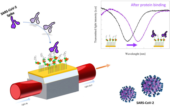
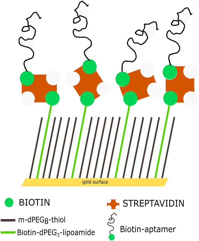
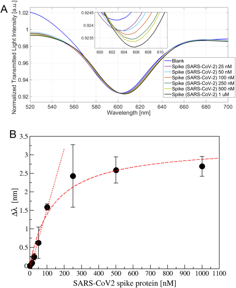
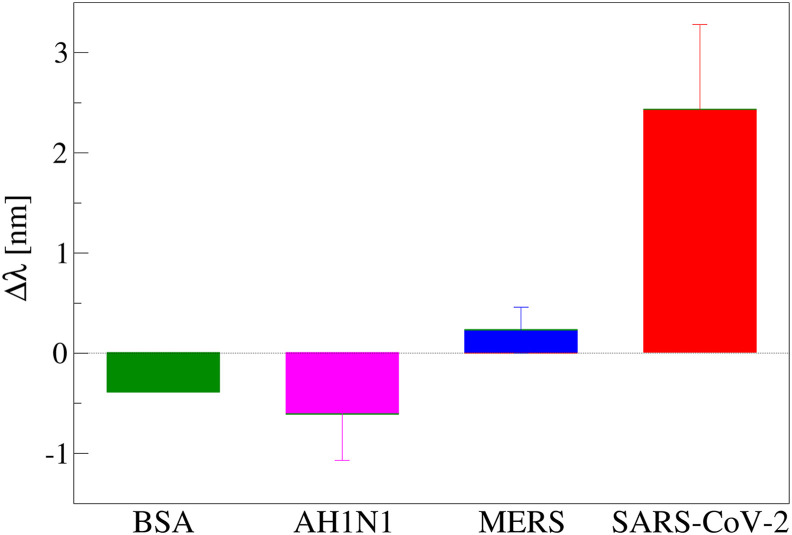
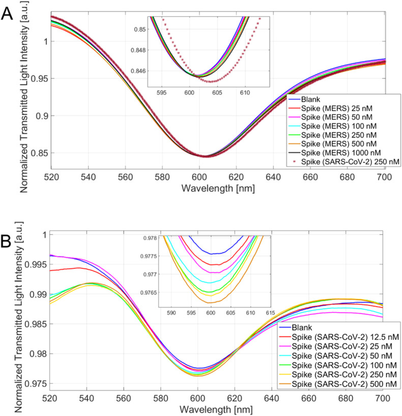
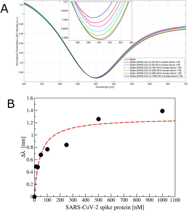
A specific aptameric sequence has been immobilized on short polyethyleneglycol (PEG) interface on gold nano-film deposited on a D-shaped plastic optical fiber (POFs) probe, and the protein binding has been monitored exploiting the very sensitive surface plasmon resonance (SPR) phenomenon. The receptor-binding domain (RBD) of the SARS-CoV-2 spike glycoprotein has been specifically used to develop an aptasensor. Surface analysis techniques coupled to fluorescence microscopy and plasmonic analysis have been utilized to characterize the biointerface. Spanning a wide protein range (25 ÷ 1000 nM), the SARS-Cov-2 spike protein was detected with a Limit of Detection (LoD) of about 37 nM. Different interferents (BSA, AH1N1 hemagglutinin protein and MERS spike protein) have been tested confirming the specificity of our aptasensor. Finally, a preliminary test in diluted human serum encouraged its application in a point-of-care device, since POF-based aptasensor represent a potentially low-cost compact biosensor, characterized by a rapid response, a small size and could be an ideal laboratory portable diagnostic tool.

Keywords: Aptamer; Plastic optical fiber (POF); Polyethyleneglycol (PEG); Sars-CoV-2; Self assembled monolayer (SAM); Surface plasmon resonance (SPR).


Conflict of interest statement

The authors declare that they have no known competing financial interests or personal relationships that could have appeared to influence the work reported in this paper.

Figures

Image 1
Graphical abstract
Scheme 1
Scheme 1
Scheme of the surface derivatization based on aptamer. A mixed layer obtained from a 0.2 mM mixture of PEGthiol and BiotinPEGlipo in 8:2 molar ratio was prepared on gold and a streptavidin coating was next performed. Finally, a biotin-modified aptamer was immobilized on the streptavidin layer.
Fig. 1
Fig. 1
A) SAM aging in different conditions at 4 °C. Fluorescence microscopy data of SAMs incubated with Cy3-SA. SAMs aged in: SAM solution (open circles), MilliQ water (black triangles up) or dried (grey squares). Data are reported as mean value of 10 images acquired on two samples. Error bars represent the standard deviation. B) Apt-THR layer aging in PBS solution at 4 °C. Data are reported as mean value of two samples. Error bars represent the standard deviation. Exposition time:0.5 s.
Fig. 2
Fig. 2
A fluorescent aptamer (apt-1C-Cy3) was immobilized on different surfaces: i) PEGthiol (mPEG), ii) PEGthiol + streptavidin (mPEG + SA), iii) PEGthiol:BiotinPEGlipo mixture in a 8:2 molar ratio (MIX) or iv) PEGlipo:BiotinPEGlipo mixture in a 8:2 molar ratio + streptavidin (MIX + SA). Three independent experiments have been performed. Five images have been acquired for each sample, reporting data as mean value and standard deviation.
Fig. 3
Fig. 3
A) Transmission spectra for different SARS-CoV-2 spike protein concentration (25 ÷ 1000 nM) with a zoom of the resonance wavelengths region (inset). B) Resonance shift (Δλ) versus the spike protein concentration (nM), measured on at least three sensor for each value. Data are reported as mean value with standard deviation. Langmuir and linear fitting are also reported.
Fig. 4
Fig. 4
Resonance wavelength variation (Δλ), with respect to the blank, obtained incubating 250 nM of different proteins (BSA, AH1H1 hemagglutinin protein, MERS spike protein and SARS spike protein) on apt-1C- modified platform (at least two platform for protein); error bars represent standard deviation.
Fig. 5
Fig. 5
A) Spectra for different MERS spike protein concentrations (25 ÷ 1000 nM) and a SARS-CoV-2 spike protein concentration (250 nM) with a zoom of the resonance wavelengths region (inset). B) Spectra for different SARS-CoV-2 spike protein concentration (12.5 ÷ 500 nM) on the apt–NS–modified platform and a zoom of the resonance wavelengths region (inset).
Fig. 6
Fig. 6
A) SPR transmission spectra, normalized to the spectrum in air, incubating different concentration of SARS-CoV-2 spike protein (25 ÷ 1000 nM) in human serum diluted 1:50 in phosphate buffer on the apt1C- aptamer functionalized POF. Inset: zoom of the resonance wavelengths region. B) Plot of the resonance shift (Δλ), with respect to the blank, versus the spike protein concentration (nM) diluted in human serum (1:50 v/v), reporting also the Langmuir fit.

References

    1. Gupta A., Kumar S., Kumar R., Choudhary A.K., Kumari K., Singh P., Kumar V. COVID-19: emergence of infectious diseases, nanotechnology aspects, challenges, and future perspectives. ChemistrySelect. 2020;5:7521–7533. doi: 10.1002/slct.202001709. - DOI - PMC - PubMed
    1. Udugama B., Kadhiresan P., Kozlowski H.N., Malekjahani A., Osborne M., Li V.Y.C., Chen H., Mubareka S., Gubbay J.B., Chan W.C.W. Diagnosing COVID-19: the disease and tools for detection. ACS Nano. 2020;14:3822–3835. doi: 10.1021/acsnano.0c02624. - DOI - PubMed
    1. Kubina R., Dziedzic A. Molecular and serological tests for COVID-19. A comparative review of SARS-CoV-2 coronavirus laboratory and point-of-care diagnostics. Diagnostics. 2020;10 doi: 10.3390/diagnostics10060434. - DOI - PMC - PubMed
    1. Malik Y.S., Kumar N., Sircar S., Kaushik R., Bhat S., Dhama K., Gupta P., Goyal K., Singh M.P., Ghoshal U., El Zowalaty M.E., Vinodhkumar O.R., Yatoo M.I., Tiwari R., Pathak M., Patel S.K., Sah R., Rodriguez-Morales A.J., Ganesh B., Kumar P., Singh R.K. Coronavirus disease pandemic (Covid-19): challenges and a global perspective. Pathogens. 2020;9:1–31. doi: 10.3390/pathogens9070519. - DOI - PMC - PubMed
    1. Piyush R., Rajarshi K., Chatterjee A., Khan R., Ray S. Nucleic acid-based therapy for coronavirus disease 2019. Heliyon. 2020;6 doi: 10.1016/j.heliyon.2020.e05007. - DOI - PMC - PubMed