. 2021 Jul 2;16(1):46.
doi: 10.1186/s13024-021-00468-x.

SETD7-mediated monomethylation is enriched on soluble Tau in Alzheimer's disease

Affiliations

SETD7-mediated monomethylation is enriched on soluble Tau in Alzheimer's disease

Maria Bichmann et al. Mol Neurodegener. .

Abstract

Background: Human tauopathies including Alzheimer's disease (AD) are characterized by alterations in the post-translational modification (PTM) pattern of Tau, which parallel the formation of insoluble Tau aggregates, neuronal dysfunction and degeneration. While PTMs on aggregated Tau have been studied in detail, much less is known about the modification patterns of soluble Tau. Furthermore, PTMs other than phosphorylation have only come into focus recently and are still understudied. Soluble Tau species are likely responsible for the spreading of pathology during disease progression and are currently being investigated as targets for immunotherapies. A better understanding of their biochemical properties is thus of high importance.

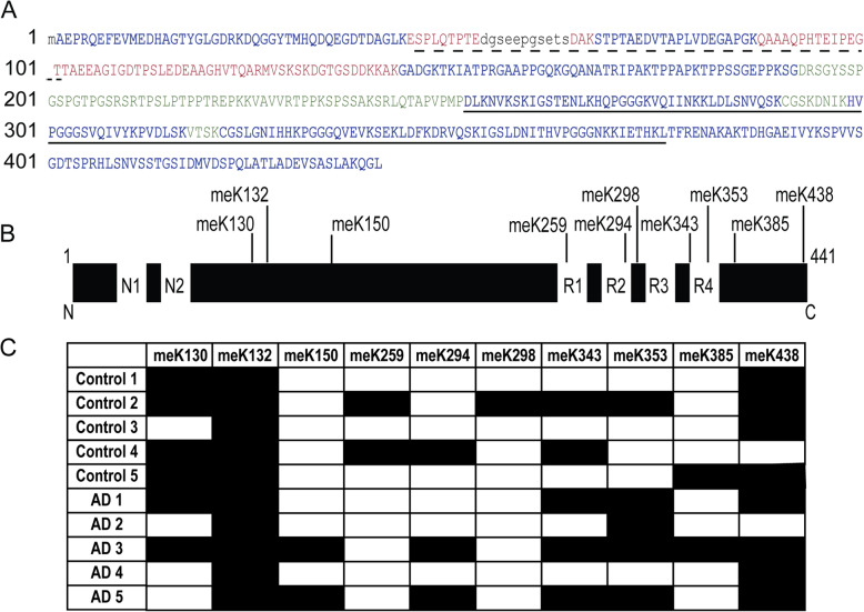
Methods: We used a mass spectrometry approach to characterize Tau PTMs on a detergent-soluble fraction of human AD and control brain tissue, which led to the discovery of novel lysine methylation events. We developed specific antibodies against Tau methylated at these sites and biochemically characterized methylated Tau species in extracts from human brain, the rTg4510 mouse model and in hiPSC-derived neurons.

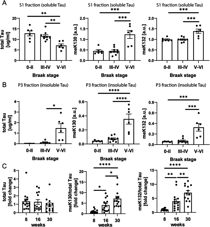
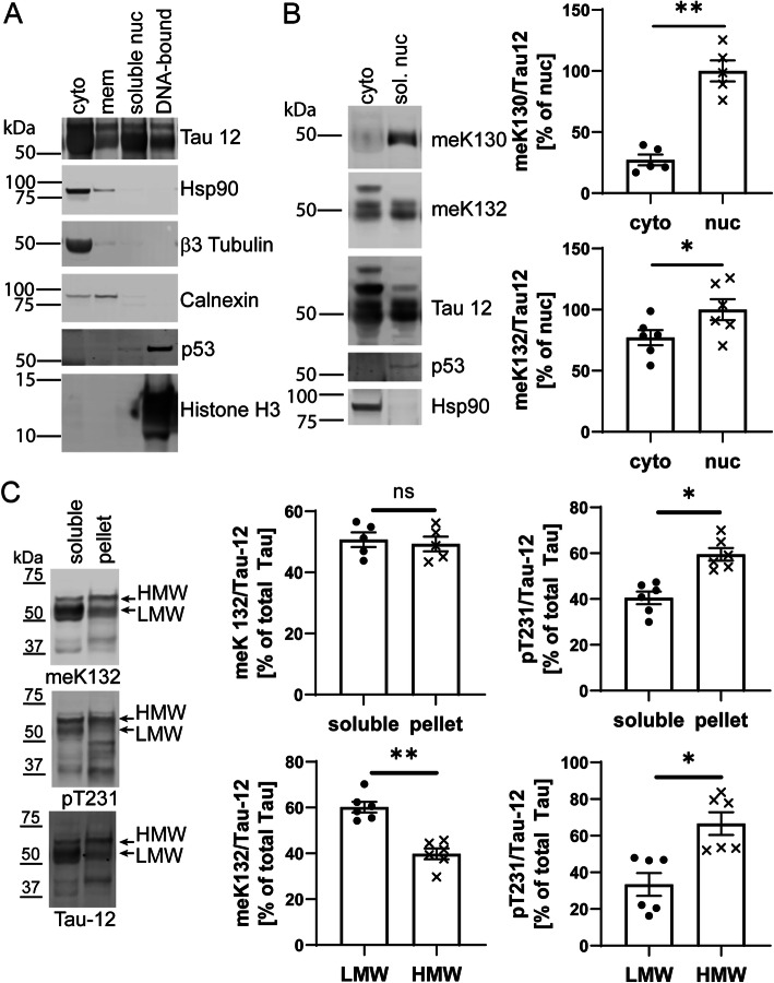
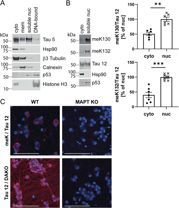
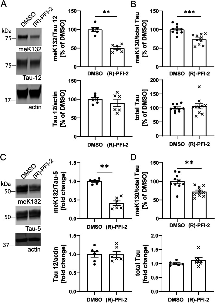
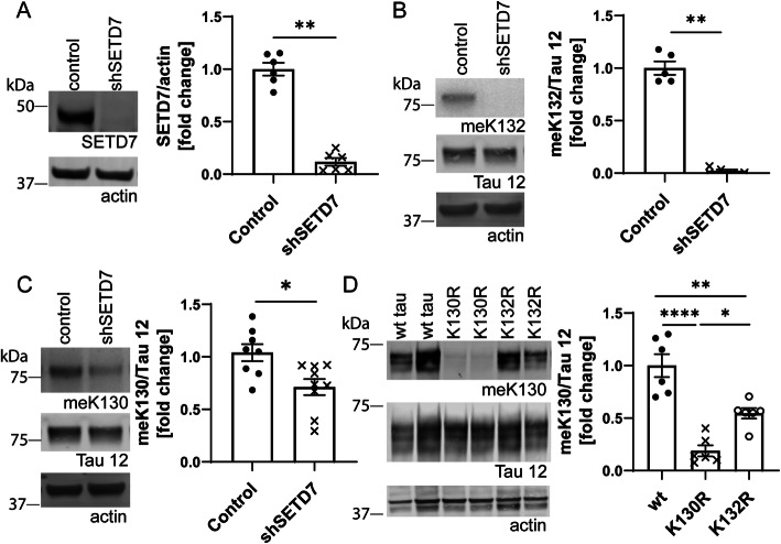
Results: Our study demonstrates that methylated Tau levels increase with Tau pathology stage in human AD samples as well as in a mouse model of Tauopathy. Methylated Tau is enriched in soluble brain extracts and is not associated with hyperphosphorylated, high molecular weight Tau species. We also show that in hiPSC-derived neurons and mouse brain, methylated Tau preferentially localizes to the cell soma and nuclear fractions and is absent from neurites. Knock down and inhibitor studies supported by proteomics data led to the identification of SETD7 as a novel lysine methyltransferase for Tau. SETD7 specifically methylates Tau at K132, an event that facilitates subsequent methylation at K130.

Conclusions: Our findings indicate that methylated Tau has a specific somatic and nuclear localization, suggesting that the methylation of soluble Tau species may provide a signal for their translocation to different subcellular compartments. Since the mislocalization and depletion of Tau from axons is associated with tauopathies, our findings may shed light onto this disease-associated phenomenon.

Keywords: Lysine methylation; Nuclear tau; Posttranslational modification; Protein methyl transferase.


Conflict of interest statement

LG, MN, AS, JSR, XW, LJ, CH and YT are employees of AbbVie, DCS and DEE joined AbbVie as employees during the study. MB, NPO, EEH, BGR, VS, PH have nothing to disclose. The financial support for this research were provided by AbbVie. AbbVie participated in the interpretation of data, review, and approval of the publication.

Figures

Fig. 1
Fig. 1
Multiple sites of lysine monomethylation detected on soluble Tau from human entorhinal cortex tissue. A Sequence coverage of the human 2N4R Tau isoform (UniProtKB-P10636–8). Dashed line: N-terminal repeats N1 and N2, solid line: microtubule binding domain (MTBR). Font color in blue: sequence covered by peptides obtained by both AspN and trypsin digestion, red: sequence covered exclusively by AspN-derived peptides, green: sequence covered exclusively by trypsin-derived peptides, black lower case: sequence not recovered by MS. B Summary of lysine monomethylation sites identified by LC-MS/MS on 2N4R Tau. C Prevalence of each monomethyl-modified lysine across the MS data set, black: found, white: not found
Fig. 2
Fig. 2
Levels of meK130 and meK132-modified Tau increase with Braak stage in humans and with age in rTg4510 mice in the soluble fraction. A Total Tau levels decrease while methylated Tau levels increase with Braak stage in the soluble fraction (S1) of human brain. B Total and methylated Tau levels increase in parallel at high Braak stages in the sarkosyl-insoluble fraction (P3). C Electrochemiluminescence ELISA demonstrates an age-dependent increase in meK130 and meK132-modified Tau in Triton-X soluble fractions of rTg4510 mouse hippocampus. Data are normalized to total Tau levels obtained by Tau 5 ELISA. A-C Statistical significance was determined by one-way ANOVA (S1 total Tau: ANOVA p < 0.001, S1 meK130: ANOVA p < 0.0001, S1 meK132: ANOVA p < 0.001; P3 total Tau: ANOVA p < 0.05, P3 meK130: ANOVA p < 0.0001, P3 meK132: ANOVA p < 0.001, rTg4510 meK130: ANOVA p = 0.0002, rTg4510 meK132: ANOVA p < 0.0001). Tukey’s multiple comparisons test was used for post-hoc analysis. *: p < 0.05, **: p < 0.01, ***: p < 0.001, ****: p < 0.0001
Fig. 3
Fig. 3
Nuclear, low-molecular weight Tau is preferentially methylated. A Cortical tissue from 30 week old rTg4510 mice was subjected to subcellular fractionation and the distribution of Tau was analyzed by Western blotting. The purity of fractions was determined by analyzing the distribution of marker proteins. As demonstrated by a representative blot, the soluble nuclear fraction contains abundant Tau protein but is free from the cytosolic proteins Hsp90, β3-Tubulin and Calnexin. B Representative Western blots and quantification demonstrate that meK130 and meK132-modified Tau preferentially localizes to the soluble nuclear fraction in the cortex of 30 week-old rTg4510 mice. C Triton-X soluble and insoluble fractions from hippocampi of 30 week-old rTg4510 mice were analyzed by Western blotting. For the distribution between the soluble and the pellet fraction, both HMW and LMW bands were quantified. While pT231-modified Tau is more abundant in the pellet and the HMW fraction, meK132-modified Tau is evenly distributed between soluble fraction and pellet and more abundant on LMW Tau. Statistical significance in (B) and (C) was determined by paired two-tailed Student’s t-test. *: p < 0.05, **: p < 0.01
Fig. 4
Fig. 4
Methylated Tau preferentially localizes to the nucleus. A hiPSC-derived neurons were subjected to subcellular fractionation and the distribution of Tau was analyzed by Western blotting. The purity of fractions was determined by analyzing the distribution of marker proteins. Similar to in vivo data, the soluble nuclear fraction contains abundant Tau protein but is free from the cytosolic protein Hsp90. B Representative Western blots and quantification demonstrate that meK130 and meK132-modified Tau preferentially localize to the soluble nuclear fraction in hiPSC-derived neurons. C hiPSC-derived neurons were subjected to proximity-ligation assays (PLA) using either a total methyl-lysine antibody (meK) in combination with Tau 12, or two total Tau antibodies (Tau 12 and DAKO). Samples were imaged on a confocal microscope. MAPT WT neurons show somatic and nuclear localization of methylated Tau, with additional neuritic staining only observed with the total Tau antibody pair. No signal was observed in MAPT KO neurons. Scale bars: 100 μm. Statistical significance in (B) was determined by paired two-tailed Student’s t-test. **: p < 0.01, ***: p < 0.001
Fig. 5
Fig. 5
The SETD7 inhibitor (R)-PFI-2 reduces Tau methylation at K130 and K132. A Representative Western blots and quantification demonstrate a significant decrease in meK132-modified Tau after treatment of SHSY5Y cells with 5 μM (R)-PFI-2 for 24 h. Total Tau levels remain unaltered. B Electrochemiluminescence ELISA shows that treatment with 5 μM (R)-PFI-2 for 24 h reduces the levels of meK130-modified Tau in SH-SY5Y cells. Total Tau levels remain unaltered. C Western blots and quantification demonstrate a significant decrease in meK132-modified Tau after treatment of hiPSC-derived neurons with 5 μM (R)-PFI-2 for 48 h. Total Tau levels remain unaltered. D Electrochemiluminescence ELISA shows that treatment with 5 μM (R)-PFI-2 for 48 h reduces the levels of meK130 Tau in iPS-derived neurons. Total Tau levels remain unaltered. Statistical significance for all panels was determined by Mann Whitney test. **: p < 0.01, ***: p < 0.001
Fig. 6
Fig. 6
shRNA-mediated knock down of SETD7 expression reduces meK130 and mek132-modified Tau in SHSY5Y cells. A Representative Western blots and quantification demonstrate a strong reduction of SETD7 protein in SHSY5Y cells treated with shRNA. B meK132-modified Tau is absent in SH-SY5Y cells treated with shRNA against SETD7, representative Western blots and quantification. C Representative Western blots and quantification demonstrate that shRNA against SETD7 also reduces the levels of meK130-modified Tau in SH-SY5Y cells, albeit to a smaller degree than the meK132 modification. D Transient transfection of WT, K130R and K132R mutant Tau 2N4R into HEK cells demonstrates that the K132R mutation significantly decreases meK130-modified Tau levels. Representative Western blots and quantification are shown. Statistical significance in (A), (B) and (C) was determined by Mann Whitney test, **: p < 0.01. Statistical significance in (D) was determined by one-way ANOVA (ANOVA p < 0.0001), Tukey’s multiple comparisons test was used for post-hoc analysis. *: p < 0.05, **: p < 0.01, ****: p < 0.0001

References

    1. Ittner A, Ittner LM. Dendritic tau in Alzheimer's disease. Neuron. 2018;99(1):13–27. doi: 10.1016/j.neuron.2018.06.003. - DOI - PubMed
    1. Sohn PD, Tracy TE, Son HI, Zhou Y, Leite RE, Miller BL, et al. Acetylated tau destabilizes the cytoskeleton in the axon initial segment and is mislocalized to the somatodendritic compartment. Mol Neurodegener. 2016;11(1):47. doi: 10.1186/s13024-016-0109-0. - DOI - PMC - PubMed
    1. Frost B, Hemberg M, Lewis J, Feany MB. Tau promotes neurodegeneration through global chromatin relaxation. Nat Neurosci. 2014;17(3):357–366. doi: 10.1038/nn.3639. - DOI - PMC - PubMed
    1. Maina MB, Bailey LJ, Wagih S, Biasetti L, Pollack SJ, Quinn JP, et al. The involvement of tau in nucleolar transcription and the stress response. Acta Neuropathol Commun. 2018;6(1):70. doi: 10.1186/s40478-018-0565-6. - DOI - PMC - PubMed
    1. Sotiropoulos I, Galas MC, Silva JM, Skoulakis E, Wegmann S, Maina MB, et al. Atypical, non-standard functions of the microtubule associated tau protein. Acta Neuropathol Commun. 2017;5(1):91. doi: 10.1186/s40478-017-0489-6. - DOI - PMC - PubMed

Publication types