. 2021 Jul;9(7):e002123.
doi: 10.1136/jitc-2020-002123.

Determinants of response to daratumumab in Epstein-Barr virus-positive natural killer and T-cell lymphoma

Affiliations

Determinants of response to daratumumab in Epstein-Barr virus-positive natural killer and T-cell lymphoma

Nurulhuda Mustafa et al. J Immunother Cancer. 2021 Jul.

Abstract

Background: The potential therapeutic efficacy of daratumumab in natural killer T-cell lymphoma (NKTL) was highlighted when its off-label usage produced sustained remission in a patient with highly refractory disease. This is corroborated recently by a phase II clinical trial which established that daratumumab monotherapy is well tolerated and displayed encouraging response in relapsed/refractory NKTL patients. However, little is known regarding the molecular factors central to the induction and regulation of the daratumumab-mediated antitumor response in NKTL.

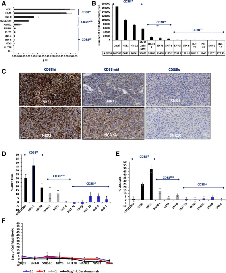
Methods: CD38 expression was studied via immunohistochemistry, multiplex immunofluorescence and correlated with clinical characteristics of the patient. The therapeutic efficacy of daratumumab was studied in vitro via CellTiter-Glo (CTG) assay, complement-dependent cytotoxicity (CDC), antibody-dependent cell cytotoxicity (ADCC), and in vivo, via a patient-derived xenograft mouse model of NKTL, both as a single agent and in combination with L-asparaginase. Signaling mechanisms were characterized via pharmacologic treatment, RNA silencing, flow cytometry and corroborated with public transcriptomic data of NKTL.

Results: Epstein-Barr virus-positive NKTL patients significantly express CD38 with half exhibiting high expression. Daratumumab effectively triggers Fc-mediated ADCC and CDC in a CD38-dependent manner. Importantly, daratumumab monotherapy and combination therapy with L-asparaginase significantly suppresses tumor progression in vivo. Ablation of complement inhibitory proteins (CIP) demonstrate that CD55 and CD59, not CD46, are critical for the induction of CDC. Notably, CD55 and CD59 expression were significantly elevated in the late stages of NKTL. Increasing the CD38:CIP ratio through sequential CIP knockdown, followed by CD38 upregulation via All-Trans Retinoic Acid treatment, potently augments complement-mediated lysis in cells previously resistant to daratumumab. The CD38:CIP ratio consistently demonstrates a statistically superior correlation to antitumor efficacy of daratumumab than CD38 or CIP expression alone.

Conclusion: This study characterizes CD38 as an effective target for a subset of NKTL patients and the utilization of the CD38:CIP ratio as a more robust identifier for patient stratification and personalisation of treatment. Furthermore, elucidation of factors which sensitize the complement-mediated response provides an alternative approach toward optimizing therapeutic efficacy of daratumumab where CDC remains a known limiting factor. Altogether, these results propose a strategic rationale for further evaluation of single or combined daratumumab treatment in the clinic for NKTL.

Keywords: antibodies; drug evaluation; hematologic neoplasms; immunotherapy; neoplasm; preclinical.


Conflict of interest statement

Competing interests: LZ and JY are staff of Janssen Pharmaceuticals.

Figures

Figure 1
Figure 1
CD38 is expressed significantly in EBV positive extranodal natural killer T-cell lymphoma (NKTL) and primary nodal NKTL (EBV +PTCL) patient samples. (A) IHC was performed in a cohort of Chinese NKTL patients (n=68, Pantonomics, California, USA) with an anti-CD38 antibody (cell Marque). To evaluate CD38 expression, membrane staining was scored as 0, 1+, 2+ or 3+, according to its intensity. H-score was then calculated by the formula (% [1+] x 1) + (% [2+] x 2) + (% [3+] x 3) as described in the Methods section. (B) This figure shows CD38/CD3/DAPI multiplexed immunofluorescence (MIF) staining on a representative panel of NKTL patient samples (n=50, NUH). Top row case shows high level of CD38 expression, with CD38+/CD3 +cells (yellow) accounting for 90% of CD3+ tumor cells (magenta). Middle row case shows medium level of CD38 staining, with CD38+/CD3 +cells (yellow) account for 30% in tumor cell population (magenta). Bottom row case represents low CD38 staining, with 0% CD38+/CD3 +cells (yellow). (L) Refers to left panel: immunofluorescence images: CD3—magenta (membrane); CD38—green (membrane); DAPI—blue (nuclear) an (R) He right panel: with corresponding marks: magenta—CD3 +cells; green—CD38+ cells; yellow—CD3+/CD38 +cells; blue—negative cells. (C) CD38 +CD3+ percentage levels from the MIF study in samples from patients diagnosed with EBV positive extranodal NKTL and primary nodal NKTL (EBV positive PTCL) were analyzed and compared. Welch T test was performed to study statistical differences. (D) The CD38 +CD3+ percentage scores were analyzed according to the staging of the cancer that patients received at diagnosis. No significant difference was observed of the CD38 +CD3+ expression between stage 2, 3 and 4 and stage 1. (E) Patient samples harvested from female and male patients were studied. Welch T test detected no significant difference between the percentage expression of CD38 +CD3+ expression in male vs female patients. (F) Kaplan-Meier analyses was performed on the overall survival between patients expressing the top 25th percentile score of CD38 +CD3+ and the lowest 25th percentile score. (G) CD38 +CD3+ percentage expression on the cell surface elucidated from multiplex immunofluorescence was correlated with CD38 gene expression levels from a microarray analyses published previously and performed on the same patient samples. The percentage surface expression of CD38+CD3+ demonstrated significant correlation with mRNA expression (Graphpad prism), where *p<0.05. EBV, Epstein-Barr virus; IHC, immunohistochemistry; NUH, National University Hospital; PTCL, peripheral T-cell lymphoma.
Figure 2
Figure 2
Daratumumab promotes anticancer activity in EBV-positive NK and T cell lymphomas through the induction of antibody-dependent cell cytotoxicity (ADCC) and complement dependent cytotoxicity (CDC). (A) Basal CD38 mRNA expression levels were studied in a panel of NKTL cell lines and one MM cell line (KMS12BM) by qRT-PCR. (B) CD38 (Pharmingen) expression on the surface of the cell lines were detected by FACS where at least 10 000 events were collected. The fluorescence of Quantibrite-PE beads (Pharmingen) was measured in parallel to create a standard curve for the elucidation of number of molecules expressed on surface of the cell. Cells were categorized into CD38hi, CD38mid and CD38lo cell lines as described in results. (C) CD38 expression on NKTL cell lines were evaluated via IHC and scored as described in 1A. (D) NKTL cell lines were preincubated with 10 µg/mL of Daratumumab or IgG. Subsequently, NKTL was cocultured with healthy donor effector primary NK cells at an effector:target ratio of 4:1. Cell lysis was measured 4 hour later (CytoTox-One, Promega). (E) NKTL cell lines were preincubated with 10 µg/mL of Daratumumab or IgG and then cultured with 20% human complement serum. Two hours later, cell viability was assessed (CTG, Promega). (F) NKTL cell lines were treated at the indicated concentrations of daratumumab for 48 hours and cell viability measured by Cell-Titer Glo (Promega). All experiments were performed at least n=3. EBV, Epstein-Barr virus; IHC, immunohistochemistry; MM, multiple myeloma; NK, natural killer; NKTL, natural killer T-cell lymphoma.
Figure 3
Figure 3
All-trans retinoic acid (ATRA) enhances anti-NKTL activity of Daratumumab by enhancing ADCC and CDC through upregulation of CD38. (A) NKTL cell lines were treated with varying doses of atra for 48 hours and cell viability (CTG, Promega) assay was performed. (B) qPCR analysis was performed in NKTL cell lines 18 hours after treatment with atra. **P<0.01 (C) CD38 cell surface expression was assessed 24 hours and 48 hours after treatment with 100 nM atra and analysis performed by FACS. At least 10 000 events were studied. *P<0.05, **p<0.01. (D, E) NKTL cell lines were treated with 100 nM of atra at the stated timepoints and (D) ADCC and (E) CDC analyzed as described previously *p<0.05, **p<0.01. (F) NKTL was treated with panobinostat for 48 hours and analyzed for CD38 surface expression by FACS. At least 10 000 events were collected. (G) NKTL was pretreated with panobinostat for 48 hours and subsequently incubated with 10 µg/mL of Daratumumab or IgG. Twenty per cent complement human serum was then added and viability of cells measured after 2 hours. All experiments were performed at least n=3. ADCC, antibody-dependent cell cytotoxicity; ATRA, all-trans retinoic acid; CDC, complement-dependent cytotoxicity; NKTL, natural killer T-cell lymphoma.
Figure 4
Figure 4
Complement inhibitory proteins (CIP) CD55 and CD59, can suppress the potency of Daratumumab-induced CDC in NKTL and are significantly associated with the later stage of this malignancy. (A) NKTL cell lines were analyzed for surface expression of membrane CIPs CD55, CD59 and CD46 by FACS and Quantibrite beads (BD) were concurrently used to identify number of molecules per cell. (B) A comparison of the IHC score of NKTL cell lines and CDC lysis induced suggests that CIPs CD55 and CD59 may be involved in regulating daratumumab-mediated CDC. (C–E) CIPs in indicated cell lines were silenced with Si Non Targeting Control (NTC) and (C) siCD46 (D) siCD55 (E) siCD55+siCD59 via electroporation (NEON). After 72 hours, CIP surface expression was detected by FACS and the effect on Daratumumab mediated CDC measured as described in Methods. **P<0.01. All experiments were performed at least n=3. (F) The ratio of CD38:CD59 expression was analyzed in NKS1 and KMS12BM before and after CD59 knockdown. The y axis on the left denotes the CD38:CD59 ratio in NKS1 and the y-axis on the right denotes the CD38:CD59 ratio of KMS12BM. The increase in CD38:CD59 ratio after CD59 knockdown was significant. *P<0.05. (G) CD55 and (H) CD59 GEP levels extracted from GSE90784 database was plotted according to the stage of cancer the patient was diagnosed, *p<0.05, **p<0.01. CDC, complement-dependent cytotoxicity; IHC, immunohistochemistry; NKTL, natural killer T-cell lymphoma; ns, not significant.
Figure 5
Figure 5
Sequential knockdown of CD55 or CD59 followed by the addition of ATRA potently enhances daratumumab-mediated cell lysis through the increase of the CD38:CIP ratio. (A) NKTL cell lines were pretreated with 100 nM atra and expression levels of CD38, CD55 and CD59 measured by FACS. At least 10 000 events were collected. Fold difference of the expression before and after atra treatment was plotted accordingly. (B) CD55 in NKYS cell lines were silenced by electroporation with siRNA for 72 hours. 24 hours after CD55 knockdown, NKYS was treated with 0 nM or 100 nM atra for the next 48 hours. subsequently, cells were harvested and analyzed for Daratumumab mediated CDC lysis. (C, D) CD55 and CD59 in (C) NKS1 cells and (D) HuT78 cells were silenced by electroporation of siRNA for 72 hours. Twenty-four hours after CD55/CD59 knockdown, cells were treated with 0 nM or 100 nM atra for the next 48 hours. After this, cells were harvested and analyzed for Daratumumab mediated CDC lysis. **P<0.01. All experiments were performed at least n=3. (E) The Spearman’s rank correlation coefficient was evaluated between CDC lysis induced and number of molecules of CD55/CD59/CD46/CD38 in the cell surface. A significant and positive correlation was observed with CD38 and a significant negative correlation associated with CD55 expression levels. Conversely, no significant correlation was observed with CD46 and CD59. (F) All the ratios of CD38:CIP that is CD38:CD46, CD38:CD55 and CD38:CD59 expression display not only a significant but also a high correlation coefficient with CDC lysis. (Graphpad Prism). ATRA, all-trans retinoic acid; CDC, complement-dependent cytotoxicity; CIP, complement inhibitory proteins; NKTL, natural killer T-cell lymphoma.
Figure 6
Figure 6
Daratumumab significantly inhibits tumor progression in a patient-derived NKTL tumor xenograft model both as a single agent and in combination with L-Asparaginase. (A) Scid beige mice were injected with patient-derived NKS1 cells and when tumors were palpable, mice were divided into three treatment groups where each group is n=10. Mice were treated with 10 mg/kg of IgG, 5 mg/kg and 10 mg/kg of daratumumab, i.p. Tumor volume was measured over time. **P<0.01. (B) Viability of the mice with Daratumumab treatment were monitored up to 30 days and plotted as a survival curve (C) mice which developed exponential tumor growth with Daratumumab treatment at the end of 30 days, ‘R’ (n=3) and mice with almost complete regression of tumor growth with Daratumumab (‘S’) were analyzed more closely. these tumors were harvested and mRNA extracted to perform a qPCR analysis for CD38, CD55, CD59 and CD46 expression. (D) NKS1 and NK92 were treated simultaneously with indicated concentrations of L-asparaginase and 3 µg/mL Daratumumab over a 24-hour period and then complement serum added and cell lysis measured by CTG (Promega). (E) NKS1 cell line was treated at IC25 and IC50 concentrations for 24 hours and the surface expression levels of CD38, CD59 and CD55 measured by FACS. At least 10 000 events were collected. (F) CB17 mice (C.B-Igh-1b/IcrTac-Prkdcscid) were injected with patient-derived NKS1 cells and when tumors were palpable, mice were divided into respective treatment groups where each group consist of n=10 mice and the mice were treated with L-Aspa 5 U/g, DarA 0.5 mg/kg+5 U/g L-Aspa, DarA 2 mg/kg+5 U/g L-Aspa, i.p. tumor volume was measured over time. **P<0.01, *p<0.05. NKTL, natural killer T-cell lymphoma.

References

    1. Villwock JA, Jones K, Back J, et al. . Delayed diagnosis of nasal natural killer/T-cell lymphoma. Case Rep Otolaryngol 2013;2013:1–4. 10.1155/2013/918132 - DOI - PMC - PubMed
    1. Yamaguchi M, Kwong Y-L, Kim WS, et al. . Phase II study of SMILE chemotherapy for newly diagnosed stage IV, relapsed, or refractory extranodal natural killer (NK)/T-cell lymphoma, nasal type: the NK-Cell Tumor Study Group study. J Clin Oncol 2011;29:4410–6. 10.1200/JCO.2011.35.6287 - DOI - PubMed
    1. Fox CP, Civallero M, Ko Y-H, et al. . Survival outcomes of patients with extranodal natural-killer T-cell lymphoma: a prospective cohort study from the International T-cell project. Lancet Haematol 2020;7:e284–94. 10.1016/S2352-3026(19)30283-2 - DOI - PubMed
    1. Haverkos BM, Pan Z, Gru AA, et al. . Extranodal NK/T cell lymphoma, nasal type (ENKTL-NT): an update on epidemiology, clinical presentation, and natural history in North American and European cases. Curr Hematol Malig Rep 2016;11:514–27. 10.1007/s11899-016-0355-9 - DOI - PMC - PubMed
    1. Qi S, Yahalom J, Hsu M, et al. . Encouraging experience in the treatment of nasal type extra-nodal NK/T-cell lymphoma in a non-Asian population. Leuk Lymphoma 2016;57:2575–83. 10.1080/10428194.2016.1180689 - DOI - PMC - PubMed

Publication types