. 2021 Jul;9(7):e002852.
doi: 10.1136/jitc-2021-002852.

STING agonist loaded lipid nanoparticles overcome anti-PD-1 resistance in melanoma lung metastasis via NK cell activation

Affiliations

STING agonist loaded lipid nanoparticles overcome anti-PD-1 resistance in melanoma lung metastasis via NK cell activation

Takashi Nakamura et al. J Immunother Cancer. 2021 Jul.

Abstract

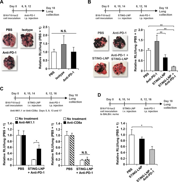
Background: Resistance to an immune checkpoint inhibitor (ICI) is a major obstacle in cancer immunotherapy. The causes of ICI resistance include major histocompatibility complex (MHC)/histocompatibility locus antigen (HLA) class I loss, neoantigen loss, and incomplete antigen presentation. Elimination by natural killer (NK) cells would be expected to be an effective strategy for the treatment of these ICI-resistant tumors. We previously demonstrated that a lipid nanoparticle containing a stimulator of an interferon gene (STING) agonist (STING-LNP) efficiently induced antitumor activity via the activation of NK cells. Thus, we evaluated the potential of reducing ICI resistance by STING-LNPs.

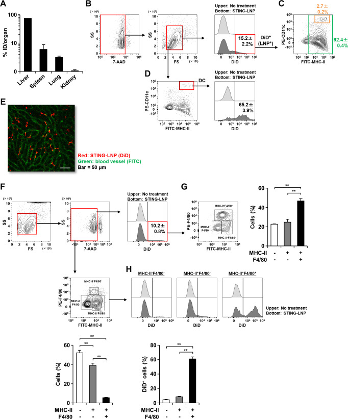
Methods: Lung metastasis of a B16-F10 mouse melanoma was used as an anti-programmed cell death 1 (anti-PD-1)-resistant mouse model. The mice were intravenously injected with the STING-LNP and the mechanism responsible for the improvement of anti-PD-1 resistance by the STING-LNPs was analyzed by RT-qPCR and flow cytometry. The dynamics of STING-LNP were also investigated.

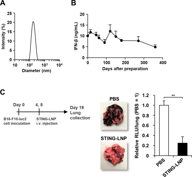
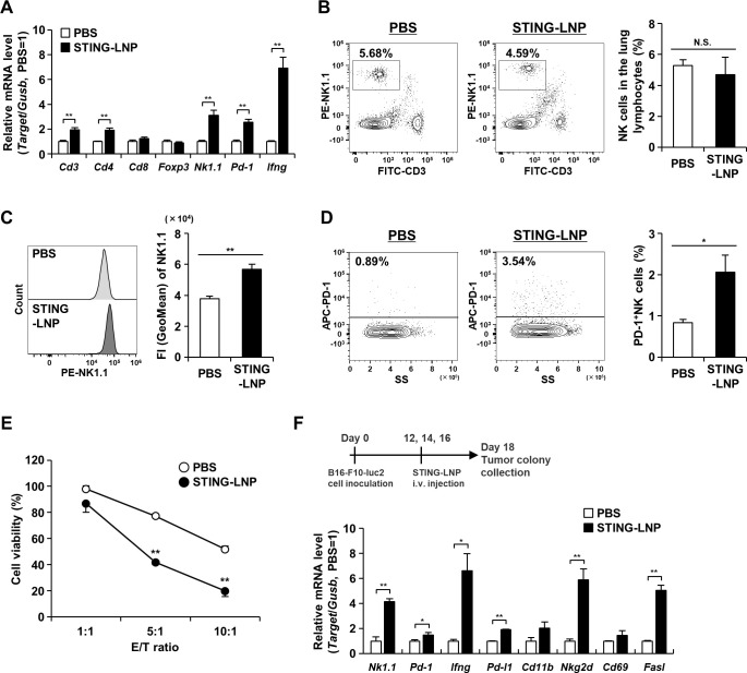
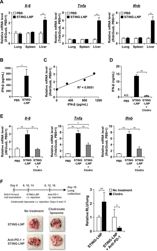
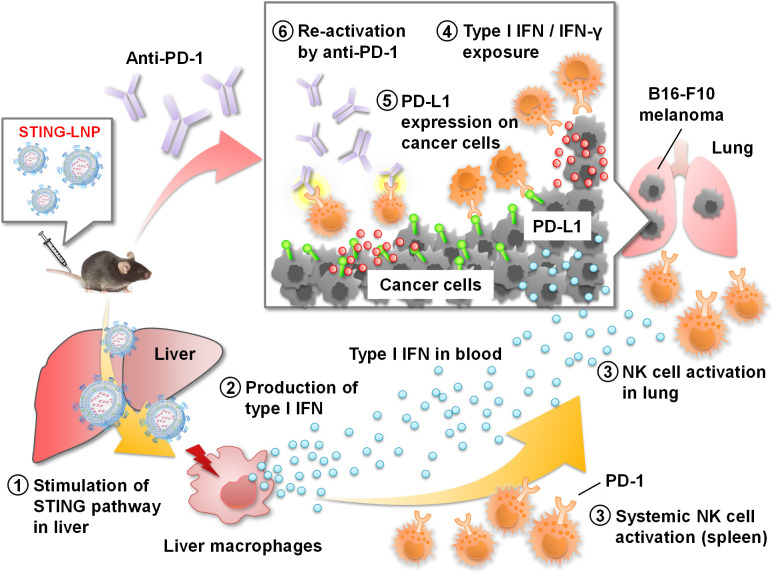
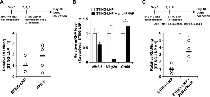
Results: Although anti-PD-1 monotherapy failed to induce an antitumor effect, the combination of the STING-LNP and anti-PD-1 exerted a synergistic antitumor effect. Our results indicate that the STING-LNP treatment significantly increased the expression of CD3, CD4, NK1.1, PD-1 and interferon (IFN)-γ in lung metastases. This change appears to be initiated by the type I IFN produced by liver macrophages that contain the internalized STING-LNPs, leading to the systemic activation of NK cells that express PD-1. The activated NK cells appeared to produce IFN-γ, resulting in an increase in the expression of the PD ligand 1 (PD-L1) in cancer cells, thus leading to a synergistic antitumor effect when anti-PD-1 is administered.

Conclusions: We provide a demonstration to show that a STING-LNP treatment can overcome PD-1 resistance in a B16-F10 lung metastasis model. The mechanism responsible for this indicates that NK cells are activated by stimulating the STING pathway which, in turn, induced the expression of PD-L1 on cancer cells. Based on the findings reported herein, the STING-LNP represents a promising candidate for use in combination therapy with anti-PD-1-resistant tumors.

Keywords: adjuvants; combination; drug therapy; immunotherapy; killer cells; melanoma; natural; pharmaceutic.


Conflict of interest statement

Competing interests: TN and HH received research funding from Ono Pharmaceutical.

Figures

Figure 1
Figure 1
Characteristics and antitumor activity of the STING-LNP. (A) Particle size distribution of STING-LNPs based on light scattering intensity. (B) Temporal change in IFN-β production in the serum by 4°C stored STING-LNP. Mice were intravenously injected with the STING-LNP which were stored at 4°C. The x-axis shows days after the preparation of STING-LNP. Data are the mean+SEM (n=3). (C) Antitumor effect of STING-LNP against B16-F10 lung metastasis. The mice were intravenously injected with the STING-LNP (6 µg of c-di-GMP). The images represent collected lungs. The value of RLU per whole lung for the PBS-treated mice were set to 1. Data are the mean+SEM (n=4, **p<0.01). c-di-GMP, cyclic di-GMP; IFN-β, interferon-β; RLU, relative light unit; STING-LNP, lipid nanoparticle containing a stimulator of an interferon gene; PBS, phosphate-buffered saline.
Figure 3
Figure 3
Analysis of lungs with B16-F10 metastasis after the STING-LNP treatment. (A) Gene expression at mRNA level in the lung with B16-F10 metastasis. Mice were intravenously injected with B16-F10-luc2 cells. The mice were intravenously injected with the STING-LNP (6 µg of c-di-GMP) on days 2, 4 and 8. On day 9, the lungs were collected, and the mRNA levels measured by RT-qPCR. The values for the PBS-treated mice were set to 1. Data are the mean+SEM (n=3, **p<0.01). (B) Analysis of NK cell population. (C) The NK1.1 expression in NK cells. (D) Analysis of PD-1+ NK cell population. B16-F10-luc2 cells were intravenously injected to mice. The mice were intravenously injected with the STING-LNP (6 µg of c-di-GMP) on day 2. On day 3, the lungs were collected, and the lymphocyte fraction obtained. The lymphocytes that were stained with anti-CD3, anti-NK1.1 and anti-PD-1 were analyzed by flow cytometry. NK cells were identified as CD3-NK1.1+ cells. Data are the mean+SEM (n=6–7, *p<0.05, **p<0.01). N.S., no significant difference. (E) Killing of B16-F10 cells by NK cells. NK cells from mice treated with PBS or the STING-LNP were mixed with B16-F10-luc2 cells at effector/target (E/T) ratio of 1:1, 5:1 and 10:1. Data are the mean+SEM (n=3, **p<0.01 vs PBS). (F) Analysis of gene expression at mRNA level in the B16-F10 colonies. The mice were intravenously injected with the STING-LNP (6 µg of c-di-GMP). The values for the PBS-treated mice were set to 1. Data are the mean+SEM (n=3, *p<0.05, **p<0.01). c-di-GMP, cyclic di-GMP; NK, natural killer; PD-1, programmed cell death 1; STING-LNP, lipid nanoparticle containing a stimulator of an interferon gene; PBS, phosphate-buffered saline.
Figure 4
Figure 4
Analysis of STING-LNP distribution. Mice were intravenously injected with the DiD-labeled STING-LNP and tissues were collected after 1 hour. (A) Biodistribution of STING-LNP. The FIs of each tissue were measured. The values represent the accumulation rate to each organ with respect to the injected dose (ID). Data are the mean+SEM (n=3). (B) Gating strategy and percentage of DiD+ cells in all splenocytes. (C) Analysis of DiD+ cells on MHC-II/CD11c plot. (D) Percentage of DiD+ cells in DCs. The splenocytes were stained with anti-I-Ab and anti-CD11c and were then analyzed by flow cytometry. DCs were identified as MHC-II+CD11c+ cells. Data are the mean±SEM (n=3). (E) CLSM image of liver. Red and green represent STING-LNP (DiD) and blood vessel (FITC), respectively. Bar=50 µm. (F) Gating strategy and percentage of DiD+ cells in liver lymphocytes. (G) Analysis of DiD+ cells on MHC-II/F4/80 plot. (H) Percentage of DiD+ cells in MHC-II-F4/80- cells, MHC-II+F4/80- cells and MHC-II+F4/80+ cells. The lymphocyte fraction was collected from the liver. After staining with anti-I-Ab and anti-F4/80, the lymphocytes were analyzed by flow cytometry. Data are the mean+SEM (n=3, **p<0.01). FI, fluorescence intensity; DC, dendritic cell; MHC-I, major histocompatibility complex class I; CLSM, confocal laser scanning microscopy; STING-LNP, lipid nanoparticle containing a stimulator of an interferon gene.
Figure 5
Figure 5
Analysis of major cells initiating innate immunity by STING-LNPs. (A) Gene expression at mRNA level. (B) IFN-β concentration in the serum. (C) Plot of relative mRNA level/IFN-β concentration. Mice were intravenously injected with B16-F10-luc2 cells. The mice were intravenously injected with the STING-LNPs (6 µg of c-di-GMP) on day 2. Tissue and blood samples were collected after 1.5 hours. The mRNA levels in each tissue and the IFN-β concentration in the serum were measured by RT-qPCR and ELISA, respectively. The values for the PBS-treated mice were set to 1 in the analysis of mRNA expression. Data are the mean+SEM (n=3, *p<0.05, **p<0.01). The relative mRNA levels of each mouse were plotted against the IFN-β concentrations for each mouse. The plot of PBS group was used the average values. (D) IFN-β concentration in the serum under macrophage depletion. (E) Gene expression at the mRNA level under macrophage depletion. Mice were intravenously injected with clodronate liposomes (Clodro). After 2 days, the mice were intravenously injected with the STING-LNPs (6 µg of c-di-GMP). After 2 hours, the blood and liver were collected. The values for the PBS-treated mice were set to 1 in the analysis of mRNA expression. Data are the mean+SEM (n=4, *p<0.05, **p<0.01). N.D., not detected. (F) The antitumor effect caused by the depletion of macrophages. Mice were then intravenously injected with the clodronate liposomes (Clodro) and the STING-LNP (6 µg of c-di-GMP). The mice were also intraperitoneally injected with 50 µg of anti-PD-1. The images represent collected lungs. The value of RLU per whole lung for the mice group that had not been treated with the clodronate liposomes (no treatment) was set to 1. Data are the mean+SEM (n=3–4, *p<0.05, **p<0.01). IFN, interferon; PD-1, programmed cell death 1; RLU, relative light unit; STING-LNP, lipid nanoparticle containing a stimulator of an interferon gene; PBS, phosphate-buffered saline.
Figure 2
Figure 2
Combination therapy against anti-PD-1-resistant B16-F10 lung metastasis. (A) Antitumor effect of anti-PD-1 monotherapy against B16-F10 lung metastasis. The mice were intraperitoneally injected with 200 µg of anti-PD-1. The images represent collected lungs. The value of RLU per whole lung for the PBS-treated mice was set to 1. Data are the mean+SEM (n=4). N.S., no significant difference. (B) Antitumor effect of combination therapy against B16-F10 lung metastasis. The mice were intravenously injected with the STING-LNP (6 µg of c-di-GMP) and were intraperitoneally injected with 50 µg of anti-PD-1. The images represent collected lungs. The value of RLU per whole lung for the PBS-treated mice was set to 1. Data are the mean+SEM (n=8–10, **p<0.01). (C) The effect of NK cell or CTL depletion on the antitumor activity by the combination therapy. Mice with B16-F10 lung metastasis were intraperitoneally injected with anti-NK1.1 (200 μg) or anti-CD8a (200 µg). The value of RLU per whole lung for the PBS-treated mice was set to 1. Data are the mean+SEM (n=3–7, *p<0.05). (D) Antitumor effect of combination therapy against B16-F10 lung metastasis in BALB/c nu/nu. The mice were intravenously injected with the STING-LNP (6 µg of c-di-GMP) and were intraperitoneally injected with 50 µg of anti-PD-1. The value of RLU per whole lung for the PBS-treated mice was set to 1. Data are the mean+SEM (n=5–6, *p<0.05). CTL, cytotoxic T lymphocyte; PD-1, programmed cell death 1; RLU, relative light unit; STING-LNP, lipid nanoparticle containing a stimulator of an interferon gene; PBS, phosphate-buffered saline.
Figure 7
Figure 7
Summary for reducing anti-PD-1 resistant by STING-LNP. (1) The intravenously injected STING-LNPs accumulate in the liver and are taken up by liver macrophages, leading to the activation of the STING pathway. (2) The liver macrophages secret type I IFNs to the blood circulation. (3) The type I IFNs activate systemic NK cells such as in the spleen and the lung. A portion of NK cells express PD-1. (4) The activated NK cells produce IFN-γ in the B16-F10 tumor. (5) The exposure of type I IFNs and IFN-γ promotes the expression of PD-L1 on the cancer cells, leading to the establishment of immunosuppression via PD-1/PD-L1 axis between PD-1+ NK cells and cancer cells. (6) An anti-PD-1 treatment blocks the immunosuppression, resulting in the reactivation of NK cells and the development of cytotoxicity against cancer cells. c-di-GMP, cyclic di-GMP; IFN, interferon; NK, natural killer; PD-1, programmed cell death 1; STING-LNP, lipid nanoparticle containing a stimulator of an interferon gene.
Figure 6
Figure 6
Effect of type I IFN on the antitumor activity by STING-LNP. (A) Comparison of the antitumor effect between the STING-LNP and the recombinant IFN-β. The mice were intravenously injected with the STING-LNP (6 µg of c-di-GMP) or recombinant IFN-β (rIFN-β) (14.6 ng). The RLU value per whole lung for the STING-LNP-treated mice was set to 1. Data are the mean+SEM (n=4–5). (B) Mice with B16-F10 lung metastasis were intraperitonially injected with 200 µg of anti-IFNAR on day 1 and intravenously injected with the STING-LNP (6 µg of c-di-GMP) on day 2 and mRNA levels were measured on day 3. The values for the STING-LNP without the anti-IFNAR group were set to 1 in the analysis for mRNA expression. Data are the mean+SEM (n=3, *p<0.05, **p<0.01). (C) Mice with B16-F10 lung metastasis were intraperitonially injected with 200 µg of anti-IFNAR and intravenously injected with the STING-LNP (6 µg of c-di-GMP). The RLU value per whole lung for the STING-LNP without anti-IFNAR group was set to 1. Data are the mean+SEM (n=4–5, **p<0.01). IFN, interferon; RLU, relative light unit; STING-LNP, lipid nanoparticle containing a stimulator of an interferon gene.

References

    1. Chen DS, Mellman I. Oncology meets immunology: the cancer-immunity cycle. Immunity 2013;39:1–10. 10.1016/j.immuni.2013.07.012 - DOI - PubMed
    1. Hildner K, Edelson BT, Purtha WE, et al. . Batf3 deficiency reveals a critical role for CD8alpha+ dendritic cells in cytotoxic T cell immunity. Science 2008;322:1097–100. 10.1126/science.1164206 - DOI - PMC - PubMed
    1. Fuertes MB, Kacha AK, Kline J, et al. . Host type I IFN signals are required for antitumor CD8+ T cell responses through CD8{alpha}+ dendritic cells. J Exp Med 2011;208:2005–16. 10.1084/jem.20101159 - DOI - PMC - PubMed
    1. Woo S-R, Fuertes MB, Corrales L, et al. . Sting-Dependent cytosolic DNA sensing mediates innate immune recognition of immunogenic tumors. Immunity 2014;41:830–42. 10.1016/j.immuni.2014.10.017 - DOI - PMC - PubMed
    1. Chen Q, Sun L, Chen ZJ. Regulation and function of the cGAS-STING pathway of cytosolic DNA sensing. Nat Immunol 2016;17:1142–9. 10.1038/ni.3558 - DOI - PubMed

Publication types