. 2021 Jul 2;11(1):13726.
doi: 10.1038/s41598-021-93143-6.

Association between overactive bladder and pelvic organ mobility as evaluated by dynamic magnetic resonance imaging

Affiliations

Association between overactive bladder and pelvic organ mobility as evaluated by dynamic magnetic resonance imaging

Kurenai Kinno et al. Sci Rep. .

Erratum in

Abstract

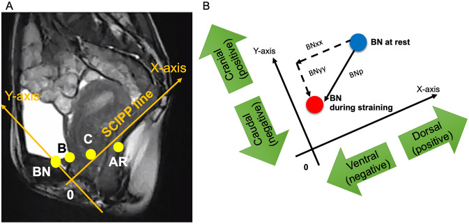
Overactive bladder (OAB) is a prevalent condition, which negatively impacts patients' quality of life. Pelvic organ prolapse (POP), also prevalent in women, has been recognized as an important etiology of female OAB, although the pathophysiological mechanisms remain controversial. In this study, we reviewed findings of dynamic magnetic resonance imaging (dMRI) in 118 patients with POP and investigated the association between dMRI findings, including positions and mobilities of pelvic organs as well as parameters of pelvic organ support and bladder outlet obstruction (urethral kinking), and OAB in order to elucidate the pathophysiology of OAB in patients with POP. Our results showed that compared with non-OAB patients, OAB patients had a significantly higher body mass index, more severe pelvic floor muscle impairment, and more profound supportive defects in the uterine cervix (apical compartment). On the other hand, dMRI parameters showed hardly any significant difference between patients with mild and moderate to severe OAB. These findings may imply that levator ani impairment and defective supports of the apical compartment could be associated with the presence of OAB and that the severity of OAB could be affected by factors other than those related to pelvic organ mobility and support or urethral kinking.


Conflict of interest statement

The authors declare no competing interests.

Figures

Figure 1
Figure 1
Graphic presentation of coordinate positions of pelvic organs at rest and during straining as well as mean values (mm) of pelvic organ mobilities during straining in patients with overactive bladder (OAB) (A) and without OAB (B). The sacrococcygeal inferior pubic point line (X-axis) and Y-axis are tilted counterclockwise based on the mean angle in relation to the bottom line on dynamic MRI images; non-OAB, 35.7° [95% confidence interval (CI) 31.3°, 37.0°]; OAB, 33.7° (95% CI 31.3°, 36.1°). To avoid further cluttering this figure, the hiatal descent and anterior vaginal wall length were not drawn on the figure. 0, inferior margin of the symphysis pubis; AR, anorectal angle; B, most dependent position of the bladder; BN, bladder neck; C, uterine cervix; H-line, length of the urogenital hiatus; iCL, imaginary line of the cardinal ligament; iUSL, imaginary line of the uterosacral ligament; X, sacrococcygeal joint.
Figure 2
Figure 2
Graphic presentation of mean values (degree) associated with bladder outlet obstruction at rest and during straining in patients with or without overactive bladder (OAB). The sacrococcygeal inferior pubic point line (X-axis) and Y-axis are tilted counterclockwise based on the mean angle in relation to the bottom line on dynamic MRI images; all patients, 35.1° [95% confidence interval (CI) 33.9°, 36.3°]. 0, inferior margin of the symphysis pubis; AUI, angle of urethral inclination BN, bladder neck; EM, external urethral meatus; PUVA, posterior urethrovesical angle.
Figure 3
Figure 3
Methods to calculate positions and mobility of pelvic organs on dynamic magnetic resonance imaging. (A) Shows the selected coordinate positions of pelvic organs in the present study, while (B) shows how to calculate pelvic organ mobility, for example mobility of BN. As shown in (B), if BN moves ventrally in parallel with the X-axis, BNxx takes a negative value, while if BN moves dorsally, BNxx takes a positive value. Likewise, if BN moves caudally in parallel with the Y-axis, BNyy takes a negative value, while if BN moves cranially, BNyy takes a positive value. AR, anorectal angle; B, most dependent position of the bladder; BN, bladder neck; C, uterine cervix; xx, distance in x-direction; yy, distance in y-direction; X-axis corresponds to the sacrococcygeal inferior pubic point (SCIPP) line and Y-axis corresponds to a perpendicular line to X-axis at the origin (0, the inferior margin of the pubic symphysis).
Figure 4
Figure 4
Methods to calculate parameters of pelvic organ support and bladder outlet obstruction on dynamic magnetic resonance imaging. (A) Shows measured parameters as follows: the anterior vaginal wall length (AVWL) is traced from the anterior vaginal fornix to the external urinary meatus; the length of the urogenital hiatus (H-line) is traced from origin to anorectal angle; the imaginary cardinal ligament (iCL) is traced from the anterior surface between the second and third sacral bones to the uterine cervix (C); the imaginary uterosacral ligament (iUSL) is traced from the anterior surface between the fourth and fifth sacral bones to C; the hiatal descent (M′-line) extends perpendicularly from the SCIPP line to the posterior end of the H-line. (B) Shows measured parameters for bladder outlet obstruction: the angle of urethral inclination (AUI) is the angle of the urethral axis in relation to the vertical plane; the posterior urethrovesical angle (PUVA) is the angle between the urethral axis and the posterior border of the bladder base or trigone.

Comment in

References

    1. Peyronnet B, et al. A comprehensive review of overactive bladder pathophysiology: On the way to tailored treatment. Eur. Urol. 2019;75:988–1000. doi: 10.1016/j.eururo.2019.02.038. - DOI - PubMed
    1. de Boer TA, et al. Pelvic organ prolapse and overactive bladder. Neurourol. Urodyn. 2010;29:30–39. doi: 10.1002/nau.20858. - DOI - PubMed
    1. Guzman-Negron J, Vasavada S. Management of overactive bladder in the face of high grade prolapse. Curr. Urol. Rep. 2017;18:12. doi: 10.1007/s11934-017-0662-2. - DOI - PubMed
    1. Obinata D, et al. Lower urinary tract symptoms in female patients with pelvic organ prolapse: Efficacy of pelvic floor reconstruction. Int. J. Urol. 2014;21:301–307. doi: 10.1111/iju.12281. - DOI - PubMed
    1. Liedl B, Goeschen K, Sutherland SE, Roovers JP, Yassouridis A. Can surgical reconstruction of vaginal and ligamentous laxity cure overactive bladder symptoms in women with pelvic organ prolapse? BJU Int. 2019;123:493–510. doi: 10.1111/bju.14453. - DOI - PubMed