. 2021 Jul 2;5(1):63.
doi: 10.1038/s41698-021-00202-2.

Validation of a liquid biopsy assay with molecular and clinical profiling of circulating tumor DNA

Affiliations

Validation of a liquid biopsy assay with molecular and clinical profiling of circulating tumor DNA

Justin D Finkle et al. NPJ Precis Oncol. .

Abstract

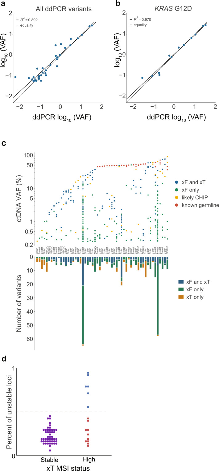
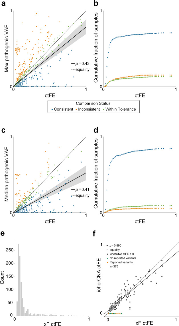
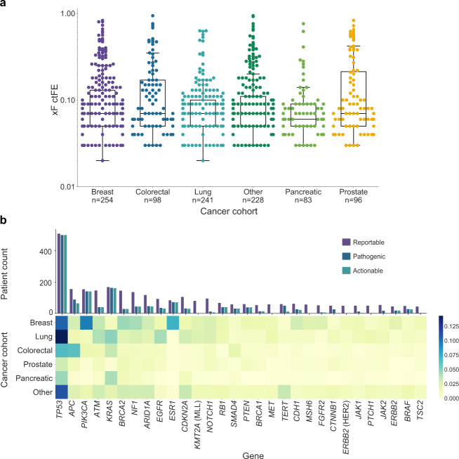
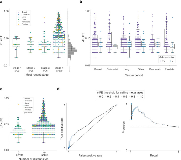
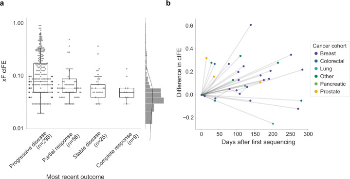
Liquid biopsy is a valuable precision oncology tool that is increasingly used as a non-invasive approach to identify biomarkers, detect resistance mutations, monitor disease burden, and identify early recurrence. The Tempus xF liquid biopsy assay is a 105-gene, hybrid-capture, next-generation sequencing (NGS) assay that detects single-nucleotide variants, insertions/deletions, copy number variants, and chromosomal rearrangements. Here, we present extensive validation studies of the xF assay using reference standards, cell lines, and patient samples that establish high sensitivity, specificity, and accuracy in variant detection. The Tempus xF assay is highly concordant with orthogonal methods, including ddPCR, tumor tissue-based NGS assays, and another commercial plasma-based NGS assay. Using matched samples, we developed a dynamic filtering method to account for germline mutations and clonal hematopoiesis, while significantly decreasing the number of false-positive variants reported. Additionally, we calculated accurate circulating tumor fraction estimates (ctFEs) using the Off-Target Tumor Estimation Routine (OTTER) algorithm for targeted-panel sequencing. In a cohort of 1,000 randomly selected cancer patients who underwent xF testing, we found that ctFEs correlated with disease burden and clinical outcomes. These results highlight the potential of serial testing to monitor treatment efficacy and disease course, providing strong support for incorporating liquid biopsy in the management of patients with advanced disease.


Conflict of interest statement

J.D.F., H.B., T.M.D., C.L., R.A.B., A.H., A.A.K., A.L., K.E.M., J.P., W.Z., K.P.W., R.T. and N.B are employees and shareholders of Tempus Labs, Inc. The remaining authors declare no competing interests.

Figures

Fig. 1
Fig. 1. Inter-assay comparison between Tempus xF, ddPCR, and Tempus xT results.
Patient samples with selected variants (n = 38) were analyzed by ddPCR and compared with xF variant allele fraction (VAF), resulting in high correlation overall (R2 = 0.892) (a), and in individual variants such as KRAS G12D (n = 12, R2 = 0.970) (b). c Results from the Tempus xF liquid biopsy and xT solid tumor assays were compared in patients who received both tests (n = 55) for colon, breast, and non-small cell lung cancers. The ctDNA VAFs for each variant are categorized by assay type in which they were detected and clonal hematopoiesis (CH) or germline status (top). The number of reportable variants for each individual patient are categorized by the assays in which they were detected (bottom). A total of 36 out of 55 xF samples had at least one pathogenic variant not detected in xT. d Among the 65 samples included in the microsatellite instability (MSI) validation cohort, 16 were deemed MSI-high and 49 microsatellite-stable. MSI was detected by xF in 6 out of 16 MSI-high patients, with 100% specificity (blue dots above dotted line).
Fig. 2
Fig. 2. Circulating tumor fraction estimate (ctFE) and variant allele fraction (VAF).
ctFE of xF-sequenced patients (n = 1000) shows a modest correlation with max pathogenic (a) and median (c) VAF (ρ = 0.43 and 0.41, respectively) even after removing likely germline variants and variants that fall within an amplified region of the genome. A variant can be detected at or below the tumor fraction of the sample, so if the detected VAF was less than the OTTER ctFE it was considered consistent (blue). If the maximum or median VAF was within ±20% of the relative OTTER ctFE, or an absolute 0.02 of the fraction, then the sample was considered within tolerance (green). If the detected VAF was greater than the OTTER ctFE it was considered inconsistent (orange). b, d Cumulative fraction of samples with consistent VAF and OTTER estimates of tumor fraction as the ctFE increases. Overall, 84.7% of samples had OTTER ctFEs within tolerance or consistent with the maximal VAF detected in the sample (b). Overall, 94.1% of samples had OTTER ctFEs within tolerance or consistent with the medial VAF detected in the sample (d). e The distribution of ctFE across the cohort (median ctFE = 0.07, mean ctFE = 0.12, SD = 0.15). f In samples that also underwent low-pass whole-genome sequencing (LPWGS, n = 375), ichorCNA detected tumor fraction in just 165 samples (black). Among those samples, there was a strong correlation between LPWGS-predicted tumor fraction and OTTER ctFE (ρ = 0.890). In the majority of samples (60%, orange) with no ctFE detected by IchorCNA, we also detected a variant using the xF assay, indicating that there was detectable tumor DNA and the ichorCNA estimate was a false negative.
Fig. 3
Fig. 3. Circulating tumor fraction estimate (ctFE) and mutational landscape by cancer type.
a Median ctFE among the most common cancer types was 0.07, with the exception of prostate (ctFE = 0.06). b The mutational landscape was evaluated in the xF 1000 cohort, with variants categorized as reportable, pathogenic, or actionable. Across all patients, the most commonly mutated gene was TP53. The heatmap was normalized within rows to depict the most prevalent variants detected for each common cancer type in the cohort (breast n = 254, colorectal n = 98, lung n = 241, prostate n = 96, and pancreatic n = 83).
Fig. 4
Fig. 4. Circulating tumor fraction estimate (cfTE) according to stage and number of distant metastases.
a Among the xF 1000 cohort, there was a significant difference in ctFE between stages (Kruskal-Wallis P=2.97e−5). Overall, patients with stage 4 cancer (n = 879, median ctFE = 0.07) had a higher ctFE than those with stages 1 (n = 20, median ctFE = 0.06), 2 (n = 25, median ctFE = 0.06), or 3 (n = 76, median ctFE = 0.06). ctFE increased with the number of metastatic distant sites (Mann-Whitney U test P = 7.57e−7) (b), and there was a significant difference in ctFEs between patients with no metastatic lesions (n = 104) and those with 1 or more distant sites affected (n = 884, Mann–Whitney U test P = 5.21e−6) (c). d ROC and PR curves for calling metastases at different ctFE thresholds. Samples with and without identified metastases (n = 100) were selected to calculate the curves with balanced classes. AUROC=0.62 and AUPR=0.64. The dashed lines show expectations when selecting samples at random.
Fig. 5
Fig. 5. Circulating tumor fraction estimate (cfTE) and abstracted clinical outcomes in a sub-cohort of the xF 1000 (n = 388).
a Patients with complete response (n = 9, ctFE = 0.05) exhibited lower ctFEs than those with progressive disease (n = 298, ctFE = 0.08), partial response (n = 56, ctFE = 0.06), or stable disease (n = 25, ctFE = 0.06). b ctFE was also assessed temporally among a few randomly selected patients with multiple xF tests throughout the course of treatment (n = 26), with most patients showing large differences in ctFEs between test dates.

References

    1. Leighl NB, et al. Clinical utility of comprehensive cell-free DNA analysis to identify genomic biomarkers in patients with newly diagnosed metastatic non-small cell lung cancer. Clin. Cancer Res. 2019;25:4691–4700. doi: 10.1158/1078-0432.CCR-19-0624. - DOI - PubMed
    1. Aggarwal C, et al. Clinical implications of plasma-based genotyping with the delivery of personalized therapy in metastatic non-small cell lung cancer. JAMA Oncol. 2019;5:173–180. doi: 10.1001/jamaoncol.2018.4305. - DOI - PMC - PubMed
    1. Kato, S. et al. Revisiting epidermal growth factor receptor (EGFR) amplification as a target for anti-EGFR therapy: analysis of cell-free circulating tumor DNA in patients with advanced malignancies. JCO Precis Oncol.10.1200/PO.18.00180 (2019). - PMC - PubMed
    1. Lee J, et al. Detection of ERBB2 (HER2) gene amplification events in cell-free DNA and response to Anti-HER2 agents in a large asian cancer patient cohort. Front Oncol. 2019;9:212. doi: 10.3389/fonc.2019.00212. - DOI - PMC - PubMed
    1. van Helden, E. J. et al. RAS and BRAF mutations in cell-free DNA are predictive for outcome of cetuximab monotherapy in patients with tissue-tested RAS wild-type advanced colorectal cancer. Mol. Oncol.10.1002/1878-0261.12550 (2019). - PMC - PubMed