. 2021 Nov;159(4):742-761.
doi: 10.1111/jnc.15462. Epub 2021 Aug 25.

Protein arginine methyltransferase 8 modulates mitochondrial bioenergetics and neuroinflammation after hypoxic stress

Affiliations

Protein arginine methyltransferase 8 modulates mitochondrial bioenergetics and neuroinflammation after hypoxic stress

Alexandre Couto E Silva et al. J Neurochem. 2021 Nov.

Abstract

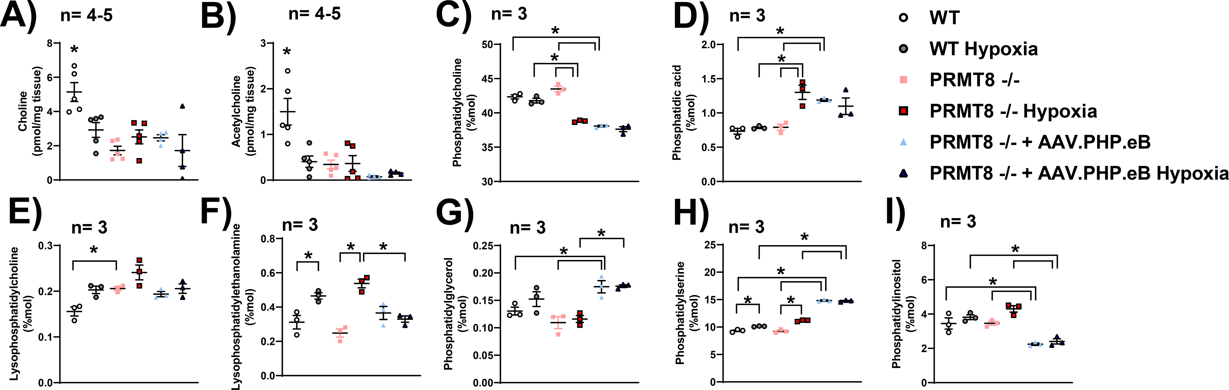
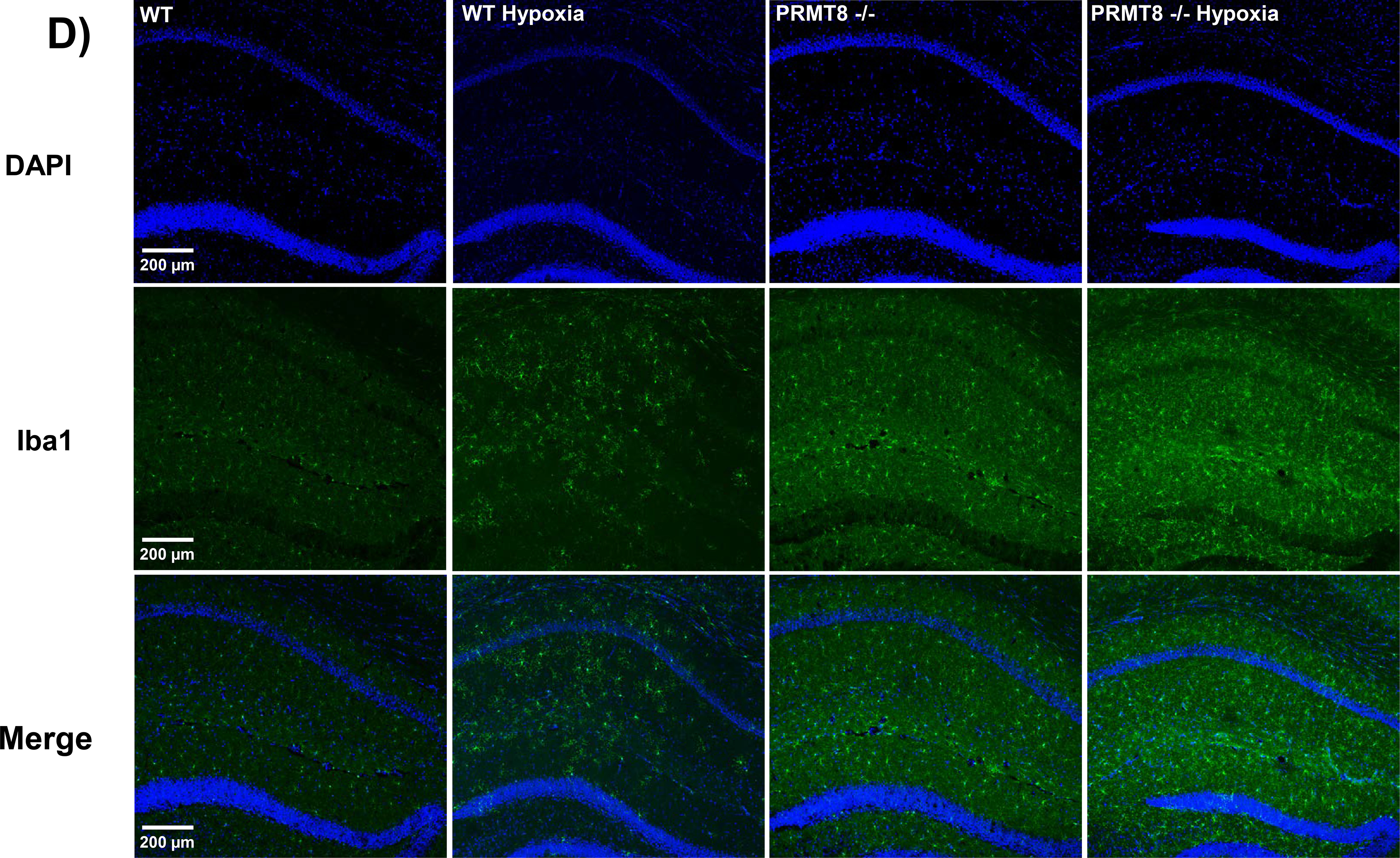
Protein arginine methyltransferases (PRMTs) are a family of enzymes involved in gene regulation and protein/histone modifications. PRMT8 is primarily expressed in the central nervous system, specifically within the cellular membrane and synaptic vesicles. Recently, PRMT8 has been described to play key roles in neuronal signaling such as a regulator of dendritic arborization, synaptic function and maturation, and neuronal differentiation and plasticity. Here, we examined the role of PRMT8 in response to hypoxia-induced stress in brain metabolism. Our results from liquid chromatography mass spectrometry, mitochondrial oxygen consumption rate, and protein analyses indicate that PRMT8(-/-) knockout mice presented with altered membrane phospholipid composition, decreased mitochondrial stress capacity, and increased neuroinflammatory markers, such as tumor necrosis factor alpha and ionized calcium binding adaptor molecule 1 (Iba1, a specific marker for microglia/macrophage activation) after hypoxic stress. Furthermore, adenovirus-based overexpression of PRMT8 reversed the changes in membrane phospholipid composition, mitochondrial stress capacity, and neuroinflammatory markers. Together, our findings establish PRMT8 as an important regulatory component of membrane phospholipid composition, short-term memory function, mitochondrial function, and neuroinflammation in response to hypoxic stress.

Keywords: hypoxic stress; mitochondrial function; neuroinflammation; phospholipids; protein arginine methyltransferase.


Conflict of interest statement

Conflict of Interest Disclosure

No conflicts of interest, financial or otherwise, are declared by the author(s).

--Human subjects --

Involves human subjects:

If yes: Informed consent & ethics approval achieved:

=> if yes, please ensure that the info “Informed consent was achieved for all subjects, and the experiments were approved by the local ethics committee.” is included in the Methods.

ARRIVE guidelines have been followed:

Yes

=> if it is a Review or Editorial, skip complete sentence => if No, include a statement in the “Conflict of interest disclosure” section: “ARRIVE guidelines were not followed for the following reason:

(edit phrasing to form a complete sentence as necessary).

=> if Yes, insert in the “Conflict of interest disclosure” section:

“All experiments were conducted in compliance with the ARRIVE guidelines.” unless it is a Review or Editorial

Figures

Figure 1.
Figure 1.. Time‐line diagram of animal experiments and endpoints.
A total number of 5 animals were excluded during our outlier assessment via the Grubbs outlier’s test (p<0.05). Moreover, one PRMT8−/− mouse was excluded during PLD1 and PLD2 mRNA analysis. One PRMT8−/− mouse treated with AAV.PHP.eB was excluded during choline/acetylcholine measurements. During open field testing, one WT hypoxic and one hypoxic PRMT8−/− treated with AAV.PHP.eB were excluded from the analysis. In addition, one PRMT8−/− mouse was excluded during our T-maze short-term memory assessment.
Figure 2.
Figure 2.. Confirmation of PRMT8−/− knockout mice.
(A) Polymerase chain-reaction (PCR) was used to determine genomic DNA from the tails of wild-type (WT) mice and PRMT8−/− (knockout) mice. “M” indicates DNA ladder with 100 base pair increments. Genotyping was confirmed via the 252 base pair product generated from PRMT8−/− mice. (B) PRMT8 mRNA reverse transcription real-time qPCR (RT-qPCR) in cortex and hippocampus of WT and PRMT8−/− mice. (C, D) Quantification from RT-qPCR of other PRMT family members in cortex (C) and hippocampus (D). Results were expressed as fold-change ± SEM; n indicates number of animals, *p ≤ 0.05 indicates significantly different from wild-type (WT) background mice (C57BL/6J), evaluated by Student’s t-test.
Figure 3.
Figure 3.. PRMT8−/− does not affect methylarginine levels, nitric oxide intermediates, and nitric oxide synthase (NOS) expression.
Mouse hippocampus were harvested after hypoxic challenge to measure arginine substrates, including asymmetric dimethylarginine (aDMA) (A), monomethylarginine (MMA) (B), symmetric dimethylarginine (sDMA) (C) on proteins. Whole-brain tissue was used to measure total nitrate+nitrite (D), nitrite (E), and nitrate (F) concentrations before and after hypoxic stress. (G) Representative images are presented as synthetic bands from capillary-based immunoassay in the mouse hippocampus. Each lane corresponds to an individual capillary electrophoresis protein sample, in which the conditions of adjacent lanes are fully independent. Neuronal NOS (nNOS) (H), endothelial NOS (eNOS) (I), and inducible NOS (iNOS) (J) protein expressions were measured and quantified via ProteinSimple ® capillary electrophoresis system. Results were expressed as mean ± SEM; n indicates number of animals, evaluated by one-way ANOVA with Tukey’s post-hoc test.
Figure 4.
Figure 4.. Overexpression of PRMT8 via AAV.PHP.eB.
Representative image of coronal (A, B) and sagittal (C, D) sections of GFP-fused PRMT8 protein 4 weeks after intravenous injection of vehicle or AAV treatment, vector genomes/kilogram (vg/kg). Cortical (E) and hippocampal (F) PRMT8 protein expressions were measured 4 weeks after intravenous administration of the PRMT8 vector (AAV-PHP.eB-hSYN1-GFP.m-PRMT8-WPRE). Results were expressed as mean ± SEM; n indicates number of animals. *p ≤ 0.05 indicates significantly different from all other groups, evaluated via one-way ANOVA with Tukey’s post-hoc test.
Figure 5.
Figure 5.. Phospholipase D (PLD) expression and activity were not changed in cortical tissue and primary cortical neurons from PRMT8−/− mice.
The hippocampus of WT and PRMT8−/− mice were analyzed with RT-qPCR for the expression of PLD1 (A) and PLD2 (B) mRNA. (C) PLD activity was measured in WT and PRMT8−/− primary cortical neuron cells via the Amplex™ Red Phospholipase D Assay Kit. Cells were treated with 10 μM of PLD inhibitor (VU0359595) as a control to establish cell/enzyme viability in both WT and PRMT8−/− cultured neurons. Results were expressed as mean ± SEM; n indicates number of animals (A,B) and independent cell culture preparations (C). *p ≤ 0.05 indicates significantly different from indicated groups, evaluated by Student’s t-test.
Figure 6.
Figure 6.. PRMT8 mediates membrane phospholipids.
(A) Choline and (B) acetylcholine (B) concentrations were measured in the cortex of WT and PRMT8−/− via a choline/acetylcholine fluorometric assay. Phosphatidylcholine (C), phosphatidic acid (D), lysophosphatidylcholine (E), lysophosphatidylethanolamine (F), phosphatidylglycerol (G), phosphatidylserine (H), and phosphatidylinositol (I) were measured by LC-MS/MS in the cortex of WT and PRMT8−/− mice. Results were expressed as mean ± SEM; n indicates number of animals. *p ≤ 0.05 indicates significantly different from indicated groups, evaluated via one-way ANOVA with Tukey’s post-hoc test.
Figure 7.
Figure 7.. Overexpression of PRMT8 restores mitochondrial respiration.
Oxygen consumption rate (OCR) was measured in hippocampal slices (200 μm) via the Seahorse XFe24 analyzer. Changes in mitochondrial respiration were manipulated by injection of 20 μg/ml oligomycin (Oligomycin), 1 mM pyruvate and 80 μM carbonyl cyanide 4-(trifluoromethoxy) phenylhydrazone (FCCP), and 20 μM antimycin A (Antimycin A) (A). Basal respiration (B), maximal respiration (C), ATP production (D), reserve capacity (E), and ATP-linked respiration (F) were measured and expressed as mean ± SEM; n indicates number of animals. *p ≤ 0.05 indicates significantly different from indicated groups, evaluated via one-way ANOVA with Tukey’s post-hoc test.
Figure 8.
Figure 8.. Overexpression of PRMT8 reduces TNF-α and microglia activation after hypoxic stress.
(A) Hippocampal protein levels of TNF-α were measured through an ELISA-based cytokine array. (B) Representative images of synthetic bands from capillary-based immunoassay. Each lane corresponds to an individual capillary electrophoresis protein sample, in which the conditions of adjacent lanes are fully independent. Quantification of ionized calcium binding adaptor molecule 1 (Iba1) expression in hippocampal proteins were summarized in C. (D) Immunohistochemistry was performed in coronal brain sections of the CA1 region of the hippocampus in WT and PRMT8−/− mice +/− hypoxia. Coronal sections were stained with Iba1 (green), a specific marker for microglia activation, and DAPI, a nuclear counterstain (blue), along with merged images (bottom row). vg/kg - vector genomes/kilogram. Results were expressed as mean ± SEM; n indicates number of animals. *p ≤ 0.05 indicates significantly different from indicated groups and respective WT backgrounds, evaluated via one-way ANOVA with Tukey’s post-hoc test.
Figure 9.
Figure 9.. PRMT8−/− mice reduced short-term memory after hypoxic stress.
(A) Short-term memory (T-maze) was assessed via spontaneous alternation protocol (Wu et al. 2018). Motor function (total distance traveled) (B) and anxiolytic behavior (time spent in center of apparatus) (C) were measured via the open field test (Feng et al. 2017). vg/kg - vector genomes/kilogram. Results were expressed as mean ± SEM; n indicates number of animals. *p ≤ 0.05 indicates significantly different from indicated groups, evaluated via one-way ANOVA with Tukey’s post-hoc test.

References

    1. Baek JH, Rubinstein M, Scheuer T and Trimmer JS (2014) Reciprocal Changes in Phosphorylation and Methylation of Mammalian Brain Sodium Channels in Response to Seizures. J Biol Chem 289, 15363–15373. - PMC - PubMed
    1. Bajwa E, Pointer CB and Klegeris A (2019) The Role of Mitochondrial Damage-Associated Molecular Patterns in Chronic Neuroinflammation. Mediators Inflamm 2019, 4050796. - PMC - PubMed
    1. Biesmans S, Bouwknecht JA, Ver Donck L et al. (2015) Peripheral Administration of Tumor Necrosis Factor-Alpha Induces Neuroinflammation and Sickness but Not Depressive-Like Behavior in Mice. Biomed Res Int 2015, 716920. - PMC - PubMed
    1. Blanc RS and Richard S (2017) Arginine Methylation: The Coming of Age. Mol Cell 65, 8–24. - PubMed
    1. Blazar BR, Levy RB, Mak TW et al. (2004) CD30/CD30 Ligand (CD153) Interaction Regulates CD4+ T cell-Mediated Graft-versus-host Disease. J Immunol 173, 2933–2941. - PubMed

Publication types

MeSH terms