. 2021 Oct 5:419:126463.
doi: 10.1016/j.jhazmat.2021.126463. Epub 2021 Jun 25.

Toxicological insights of Spike fragments SARS-CoV-2 by exposure environment: A threat to aquatic health?

Affiliations

Toxicological insights of Spike fragments SARS-CoV-2 by exposure environment: A threat to aquatic health?

Ives Charlie-Silva et al. J Hazard Mater. .

Abstract

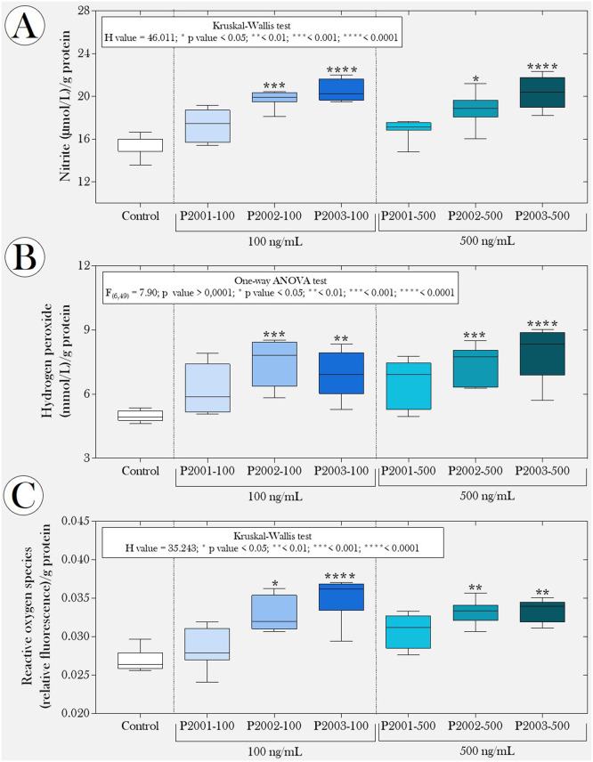
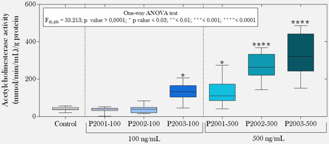
The Spike protein (S protein) is a critical component in the infection of the new coronavirus (SARS-CoV-2). The objective of this work was to evaluate whether peptides from S protein could cause negative impact in the aquatic animals. The aquatic toxicity of SARS-CoV-2 Spike protein peptides derivatives has been evaluated in tadpoles (n = 50 tadpoles/5 replicates of 10 animals) from species Physalaemus cuvieri (Leptodactylidae). After synthesis, purification, and characterization of peptides (PSDP2001, PSDP2002, PSDP2003) an aquatic contamination has been simulated with these peptides during 24 h of exposure in two concentrations (100 and 500 ng/mL). The control group ("C") was composed of tadpoles kept in polyethylene containers containing de-chlorinated water. Oxidative stress, antioxidant biomarkers and AChE activity were assessed. In both concentrations, PSPD2002 and PSPD2003 increased catalase and superoxide dismutase antioxidants enzymes activities, as well as oxidative stress (nitrite levels, hydrogen peroxide and reactive oxygen species). All three peptides also increased acetylcholinesterase activity in the highest concentration. These peptides showed molecular interactions in silico with acetylcholinesterase and antioxidant enzymes. Aquatic particle contamination of SARS-CoV-2 has cholinesterasic effect in P. cuvieri tadpoles. These findings indicate that the COVID-19 can constitute environmental impact or biological damage potential.

Keywords: Acetylcholinesterase; Amphibians; Coronavirus; Oxidative stress; SARS-Cov-2.


Conflict of interest statement

The authors declare that they have no known competing financial interests or personal relationships that could have appeared to influence the work reported in this paper.

Figures

ga1
Graphical abstract
Fig. 1
Fig. 1
Structural models of peptides (A) PSPD2001, (B) PSPD2002, and (C) PSPD2003 that were synthesized in the present study.
Fig. 2
Fig. 2
Alignment of the nucleotide sequence encoding PSPD2001, PSPD2002 and PSPD2003 with SARS-CoV-2 obtained from the (A) COVID dataset from Australia (SARS-Cov-2/human/AUS), (B) Texas, USA (SARS-Cov-2/human/TX) and (C) Iran (SARS-Cov-2/human/IRN). The blue markings refer to similar nucleotides between the synthesized peptides and those present in SARS-CoV-2. (For interpretation of the references to colour in this figure legend, the reader is referred to the web version of this article.)
Fig. 3
Fig. 3
(A) Peptide alignment and (B) Guide Tree Phylogram. Both for the Physalaemus cuvieri form (taxid: 218685) by the Clustal W program. The green regions highlight 50% of the conserved region and, in red, 50–85% of the conserved region. (For interpretation of the references to colour in this figure legend, the reader is referred to the web version of this article.)
Fig. 4
Fig. 4
Boxplot of data obtained from predictive oxidative stress biomarkers [(A) nitrite levels, (B) hydrogen peroxide and (C) reactive oxygen species] in tadpoles of P. cuvieri (phase 27 G) exposed or not to peptides PSPD 2001, 2002 and 2003 of the SARS-CoV-2 Spike protein. The summaries of the statistical analyzes are shown in the upper left corner of the graphs. Asterisks indicate significant differences between the respective groups and the control group. (n = 50 animals/group). PSPD2001: Arg-Val-Tyr-Ser-Ser-Ala-Asn-Asn-Cys- COOH; PSPD2002: Gln-Cys-Val-Asn-Leu-Thr-Thr-Arg-Thr-COOH; PSPD2003: Asn-Asn-Ala-Thr-Asn-COOH. Asterisks indicate significant differences in comparison to the control group: *p ˂ 0.05; **p ˂ 0.01; ***p < 0001; and ****p < 0,0001.
Fig. 5
Fig. 5
Boxplot of the activity of the enzymes (A) superoxide dismutase and (B) catalase, as well as correlations between the levels of (C) superoxide dismutase and (D) catalase and the different predictive biomarkers of oxidative stress. NO2-: nitrite; H2O2: hydrogen peroxide and ROS: reactive oxygen species. In "A" and "B," the statistical analyses' summaries are shown in the graphs' upper left corner. Asterisks indicate significant differences between the respective groups and the control group. (n = 50 animals/group). PSPD2001: Arg-Val-Tyr-Ser-Ser-Ala-Asn-Asn-Cys- COOH; PSPD2002: Gln-Cys-Val-Asn-Leu-Thr-Thr-Arg-Thr-COOH; PSPD2003: Asn-Asn-Ala-Thr-Asn-COOH. Asterisks indicate significant differences in comparison to the control group: *p ˂ 0.05; **p ˂ 0.01; ***p < 0001; and ****p < 0,0001.
Fig. 6
Fig. 6
Three-dimensional surface-ligand coupling of interactions between peptides PSPD2002 and PSPD-2003 and the enzyme (A-B) superoxide dismutase (SOD) and (C-D) catalase (C-D), all in surface mode and highlighted active site. In “B and D”, we also observe regions A and B of the homo-dimer structure. Interaction residues in “A” (SOD-PSPD2002): G12A; N51A *; V7A; V146B; G10B *; G12B *; N51B (affinity (kcal/mol) = −8.3). In “B” (SOD-PSPD2002): N51A; V7A; V146A; N51B **; V7B; V146B * (affinity (kcal/mol) = −8.6). In “C” (Catalase-PSPD2002): K457; N240; N451; R116**; S243 (affinity (kcal/mol) = −9.3). In “D” (Catalase-PSPD2003): D458; H455; N451; Q157*; R116 (affinity (kcal/mol) = −6.8). An "asterisk" indicates two interactions in the same residue. Two "asterisks" indicate the existence of three interactions in the same residue.
Fig. 7
Fig. 7
Boxplot of the enzyme acetylcholinesterase activity evaluated in tadpoles of P. cuvieri exposed or not to the peptides PSPD 2001, 2002, and 2003 of the SARS-CoV-2 Spike protein. The summaries of the statistical analyzes are shown in the upper left corner of the graphs. Asterisks indicate significant differences between the respective groups and the control group. (n = 50 animals/group). PSPD2001: Arg-Val-Tyr-Ser-Ser-Ala-Asn-Asn-Cys- COOH; PSPD2002: Gln-Cys-Val-Asn-Leu-Thr-Thr-Arg-Thr-COOH; PSPD2003: Asn-Asn-Ala-Thr-Asn-COOH. Asterisks indicate significant differences in comparison to the control group: *p ˂ 0.05; **p ˂ 0.01; ***p < 0001; and ****p < 0,0001.
Fig. 8
Fig. 8
Three-dimensional surface-ligand coupling of interactions between peptides (A) PSPD2002 and (B) PSPD-2003 and the enzyme acetylcholinesterase, all in surface mode and highlighted active site. Interaction residues in “A” (AChE-PSPD2002): P256; Q386*; R313**; W549 (affinity (kcal / mol) = −9.4). In “B” (AChE-PSPD2003): K434; N550; R551; V543; Y430; Y520 (affinity (kcal / mol) = −8.4). An "asterisk" indicates two interactions in the same residue. Two "asterics" indicate the existence of three interactions in the same residue.

References

    1. Abouhashem A.S., Singh K., Azzazy H.M., Sen C.K. Is low alveolar type II cell SOD3 in the lungs of elderly linked to the observed severity of COVID-19? Antioxid. Redox Signal. 2020;33:59–65. - PMC - PubMed
    1. Abu-Qdais H.A., Al-Ghazo M.A., Al-Ghazo E.M. Statistical analysis and characteristics of hospital medical waste under novel Coronavirus outbreak. Glob. J. Environ. Sci. Manag. 2020;6:21–30. Special Issue (Covid-19)
    1. Abu-Rayash A., Dincer I. Analysis of the electricity demand trends amidst the COVID-19 coronavirus pandemic. Energy Res. Soc. Sci. 2020;68 - PMC - PubMed
    1. Åkerström S., Mousavi-Jazi M., Klingström J., Leijon M., Lundkvist Å., Mirazimi A. Nitric oxide inhibitsthereplicationcycleofsevereacuterespiratorysyndrome coronavirus. J. Virol. 2005;79(3):1966–1969. - PMC - PubMed
    1. Alvarez R.A., Berra L., Gladwin M.T. Home nitric oxide therapyfor COVID-19. Am. J. Respir. Crit. Care Med. 2020;202(1):16–20. - PMC - PubMed

Publication types

Substances