. 2021 Jul 3;19(1):287.
doi: 10.1186/s12967-021-02948-6.

ALKBH3 partner ASCC3 mediates P-body formation and selective clearance of MMS-induced 1-methyladenosine and 3-methylcytosine from mRNA

Affiliations

ALKBH3 partner ASCC3 mediates P-body formation and selective clearance of MMS-induced 1-methyladenosine and 3-methylcytosine from mRNA

Kristian Lied Wollen et al. J Transl Med. .

Abstract

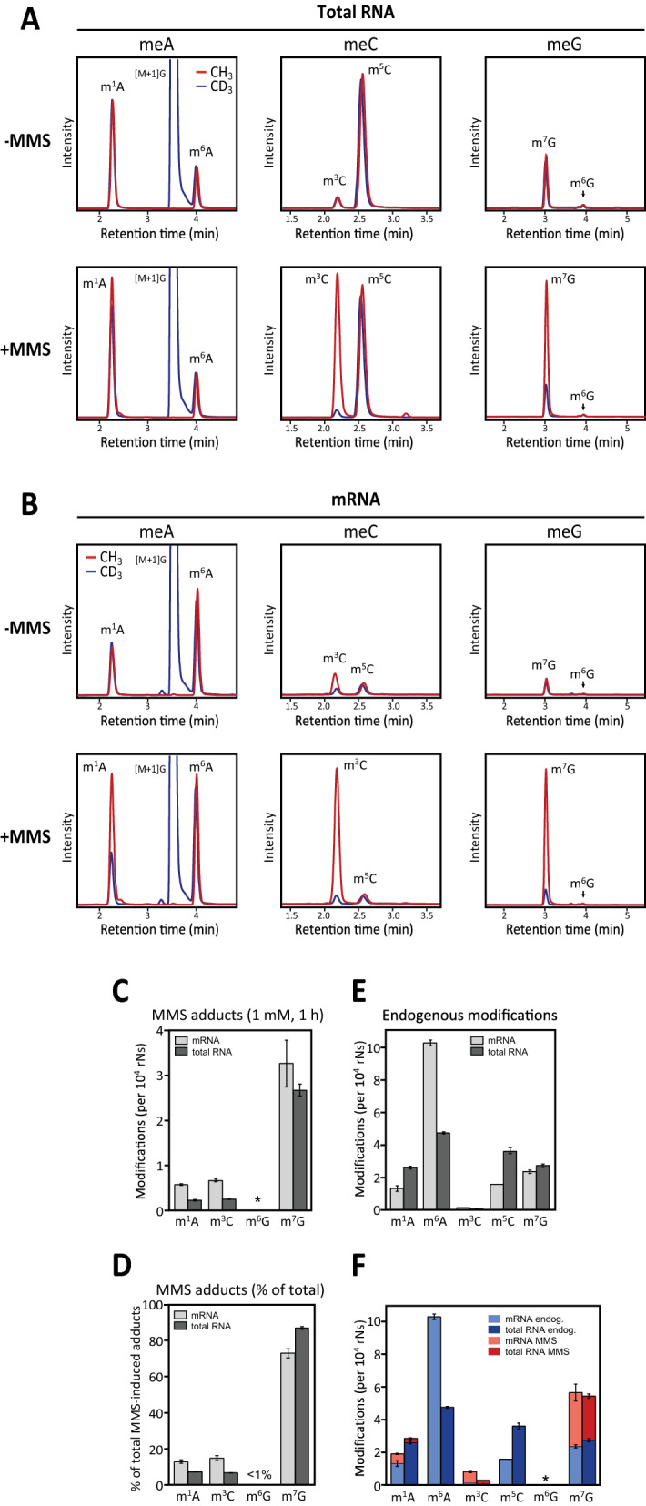
Background: Reversible enzymatic methylation of mammalian mRNA is widespread and serves crucial regulatory functions, but little is known to what degree chemical alkylators mediate overlapping modifications and whether cells distinguish aberrant from canonical methylations.

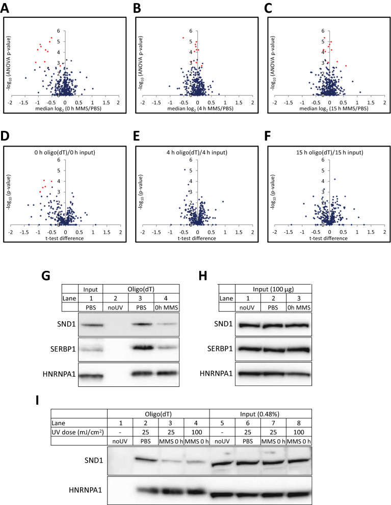
Methods: Here we use quantitative mass spectrometry to determine the fate of chemically induced methylbases in the mRNA of human cells. Concomitant alteration in the mRNA binding proteome was analyzed by SILAC mass spectrometry.

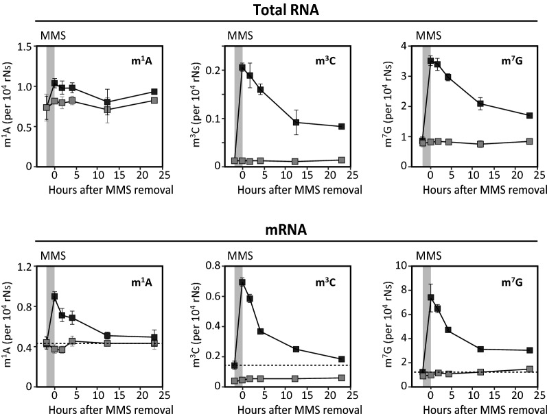
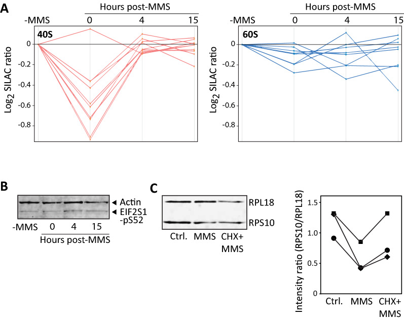
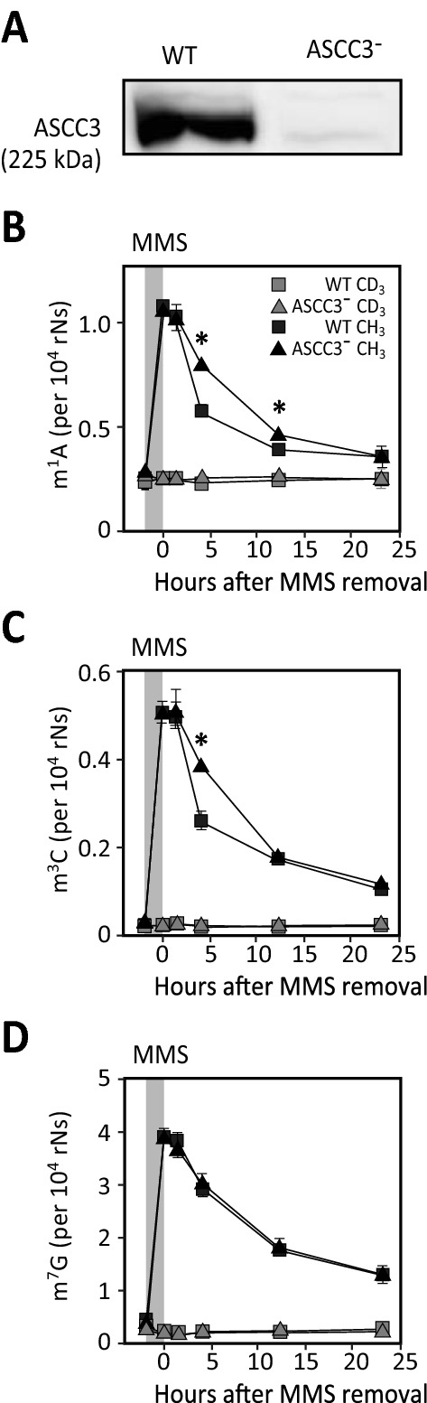
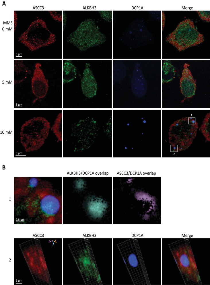
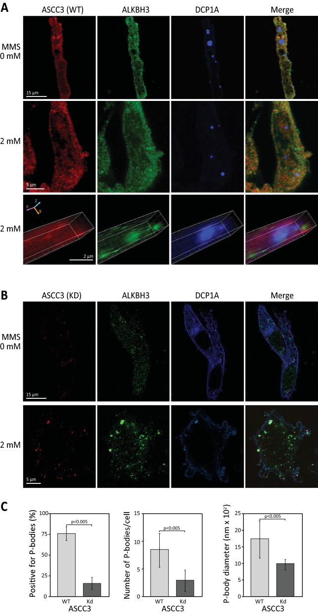
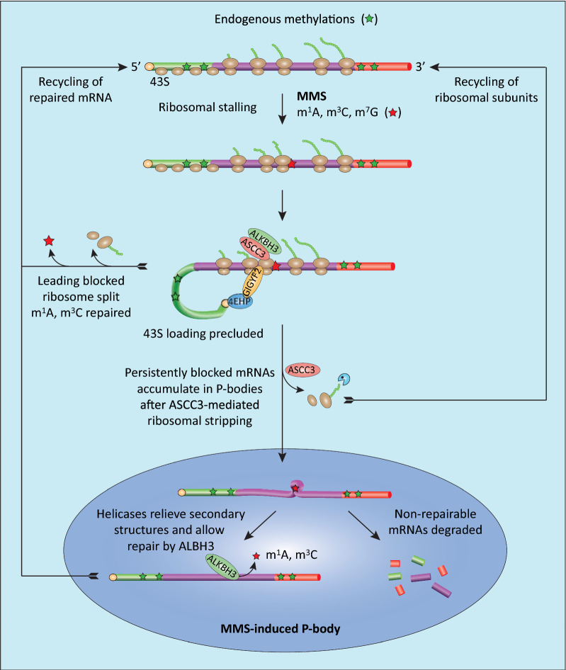
Results: MMS induced prominent direct mRNA methylations that were chemically identical to endogenous methylbases. Transient loss of 40S ribosomal proteins from isolated mRNA suggests that aberrant methylbases mediate arrested translational initiation and potentially also no-go decay of the affected mRNA. Four proteins (ASCC3, YTHDC2, TRIM25 and GEMIN5) displayed increased mRNA binding after MMS treatment. ASCC3 is a binding partner of the DNA/RNA demethylase ALKBH3 and was recently shown to promote disassembly of collided ribosomes as part of the ribosome quality control (RQC) trigger complex. We find that ASCC3-deficient cells display delayed removal of MMS-induced 1-methyladenosine (m1A) and 3-methylcytosine (m3C) from mRNA and impaired formation of MMS-induced P-bodies.

Conclusions: Our findings conform to a model in which ASCC3-mediated disassembly of collided ribosomes allows demethylation of aberrant m1A and m3C by ALKBH3. Our findings constitute first evidence of selective sanitation of aberrant mRNA methylbases over their endogenous counterparts and warrant further studies on RNA-mediated effects of chemical alkylators commonly used in the clinic.

Keywords: 1-Methyladenosine; 3-Methylcytosine; 7-Methylguanosine; ALKBH3; ASCC3; Alkylating agents; Epitranscriptome; No-go decay; P-bodies; Ribosome quality control.


Conflict of interest statement

The authors declare no conflict of interest.

Figures

Fig. 1
Fig. 1
Endogenous and MMS-induced base methylations in total- and mRNA in HeLa cells. A Mass chromatograms of methylated bases in total RNA isolated from non-treated (upper panels) or MMS-treated (lower panels) HeLa cells after 1 h exposure of 1 mM MMS. Red graphs represent fraction of the bases containing light (CH3) methyl and blue graphs represent fraction containing deuterated (CD3) methyl in each modified nucleoside. Increased CH3/CD3 ratios after MMS treatment are mediated by non-enzymatic, MMS-mediated methylation (CH3). B Similar as in A, but with mRNA. C Concentrations of MMS-induced methyl adducts in total- and mRNA. D Relative distribution of MMS-induced methyl adducts in total- and mRNA. E Endogenous levels of methylated bases in total- and mRNA in untreated cells. F Total number of various base methylations in total- and mRNA with the relative amounts of endogenous and MMS-induced base methylations as indicated by differential coloring. Each bar in C-F represents the mean of three biological replicates with SDs as indicated
Fig. 2
Fig. 2
Kinetics of removal of endogenous and MMS-induced m1A, m3C and m7G from total-and mRNA subsequent to 1 h exposure of HeLa cells to 1 mM MMS. Grey curves: endogenous (CD3) modifications; Black curves: aberrant, MMS-induced (CH3) + endogenous (CH3) modifications. Grey rectangle: period of MMS treatment. Dashed line: Baseline MMS-induced lesions
Fig. 3
Fig. 3
Temporal changes in the mRNA binding proteome after MMS treatment. AC ANOVA p-values (−log10) plotted against median SILAC ratios (log2) at 0 h (A), 4 h (B) or 15 h (C). DF Volcano plots showing t-test p-values (−log10) versus median SILAC ratios (log2) of oligo(dT)/input extract at 0 h (D), 4 h (E) and 15 h (F). Significantly altered proteins after Benjamini Hochberg FDR correction (< 0.05) are shown in red. G-I Verification of SILAC data by western analysis. HeLa cells were treated as indicated with either PBS (control) or MMS for 1 h, lysed and subjected to oligo(dT) enrichment. G SND1 and SERBP1 show reduced binding to m RNA, while HNRNPA1 does not alter its binding. H Input of extracts utilized for oligo(dT) enrichment in G. Note that the protein levels in lanes 1–3 remain the same. I Reduced mRNA binding is not caused by cross-linking bias. As in G, but MMS-treated cells were irradiated with either 25 mJ/cm2 (standard) or 100 mJ/cm2 (4 × standard dose). Note that increasing the UVC dose does not lead to increased cross-linking (lane 3 vs. lane 4)
Fig. 4
Fig. 4
Selective loss of 40S subunits from MMS-treated mRNA. A Five out of ten 40S subunits showed statistically significant transiently reduced mRNA binding after MMS treatment, whereas additional four 40S subunits demonstrated very similar, although sub-significantly altered binding pattern (left panel). Such transiently reduced binding was not observed with the 60S subunits (right panel). B 1 h MMS-treatment mediated negligible change in the level of phosphorylated (S52) EIF2S1 within 15 h after treatment (normalized to actin). C Western analysis (left panel) of mRNA-binding proteins after oligo(dT) capture confirmed a marked decrease in the ratio of bound 40S (RPS10) versus 60S (RPL18) subunits in agreement with the SILAC results. Notably, treatment with cycloheximide (10 μg/ml) for 15 min prior to the MMS treatment (CHX) to decrease ribosomal collision, partially or fully restored the RPS10/RPS18 ratio. The right panel shows RPS10/RPL18 ratios bound to mRNA in three independent experiments (given as three individual graphs) after western blotting and probing with IRDye® secondary antibodies and demonstrating the same trend (Spearman rank correlation = 1 between the three series)
Fig. 5
Fig. 5
Knockdown of ASCC3 mediates delayed removal of aberrant m1A and m3C, but not m7G, from the mRNA pool. PC-3 prostate cancer cells were subjected to stable lentiviral knockdown of ASCC3. Knockdown and wild type (WT) cells were treated with 1 mM MMS for 1 h (light grey field) and mRNA extracted at various time points. Each data point represents the mean of three biological replicates with SDs as indicated. Removal of MMS-induced (CH3) m1A (A) and m3C (B) was significantly reduced in the ASCC3-deficient cells compared to WT at 4 h and 12 h (m1A) and 4 h (m3C), respectively (* t-test, p < 0.001), whereas no such effect was observed for removal of m7G (C)
Fig. 6
Fig. 6
MMS-induced P-bodies in HeLa are enriched in ALKBH3 and localized adjacent to ASCC3. A HeLa cells transfected with P-body marker CFP-DCP1A were treated for 1 h as indicated, fixed, and double stained with anti-ASCC3 (red) and anti-ALKBH3 (green). B Enlarged images from white squares in A showing overlap of DCP1A and ALKBH3, and accumulation of ASCC3 at the P-body periphery (upper panel, corresponding to section 1). The lower panel shows z-stacks of section 2 demonstrating that ASCC3 accumulates around MMS-induced P-bodies whereas ALKBH3 accumulates inside the same P-bodies (lower panel)
Fig. 7
Fig. 7
ASCC3 promotes formation of P-bodies in PC-3 cells. PC-3 cells transfected with P-body marker CFP-DCP1A were MMS-treated for 1 h as indicated, fixed, and double stained with anti-ASCC3 (red) and anti-ALKBH3 (green). A PC-3 cells proficient in ASCC3 contain P-bodies both in the absence (upper panel) and presence (middle panel) of MMS-treatment. The bottom panel shows enlarged z-stack demonstrating co-localization of DCP1A and ALKBH3, whereas the P-body interior contains less ASCC3 than the volume immediately surrounding the same P-body. B Formation of P-bodies was severely impeded in ASCC3 knockdown PC-3 cells both in the presence and absence of MMS. C Quantitative analysis of P-bodies in WT and ASCC3 knockdown (Kd) PC-3 after 2 mM MMS treatment demonstrated that significantly fewer ASCC3-deficient cells contained P-bodies compared to WT. In the cells that contained P-bodies, these were also significantly fewer and smaller than in the parental cells
Fig. 8
Fig. 8
Working model of cellular processing of aberrantly (MMS-) methylated mRNA. MMS-induced m1A and m3C mediate direct ribosomal stalling, whereas m7G may form translation-blocking secondary structures. Collided disomes are detected by ZNF598, which serves two functions: It stabilizes GIGYF2-4EHP to block cap-dependent 43S entry and facilitates recruitment of ASCC3/ALKBH3 to mediate ribosome splitting and removal of m1A/m3C. Repaired transcripts can then be re-routed directly to translation. Transcripts harboring persistent blocking lesions, e.g. MMS-induced secondary structures, are stripped of ribosomes by ASCC3 and accumulate in P-bodies. Here, helicase activities relieve secondary structures to promote repair by ALBH3, whereas nonrepairable mRNAs are degraded

References

    1. Jones JD, Monroe J, Koutmou KS. A molecular-level perspective on the frequency, distribution, and consequences of messenger RNA modifications. Wiley Interdiscip Rev RNA. 2020;11(4):e1586. doi: 10.1002/wrna.1586. - DOI - PMC - PubMed
    1. Mathlin J, Le Pera L, Colombo T. A census and categorization method of epitranscriptomic marks. Int J Mol Sci. 2020;21(13):4684. doi: 10.3390/ijms21134684. - DOI - PMC - PubMed
    1. Tsai K, Cullen BR. Epigenetic and epitranscriptomic regulation of viral replication. Nat Rev Microbiol. 2020;18(10):559–570. doi: 10.1038/s41579-020-0382-3. - DOI - PMC - PubMed
    1. Shatkin AJ. Capping of eucaryotic mRNAs. Cell. 1976;9(4 PT 2):645–653. doi: 10.1016/0092-8674(76)90128-8. - DOI - PubMed
    1. Chu JM, Ye TT, Ma CJ, Lan MD, Liu T, Yuan BF, et al. Existence of internal N7-methylguanosine modification in mRNA determined by differential enzyme treatment coupled with mass spectrometry analysis. ACS Chem Biol. 2018;13(12):3243–3250. doi: 10.1021/acschembio.7b00906. - DOI - PubMed

Publication types