. 2021 Jul 3;14(1):105.
doi: 10.1186/s13041-021-00819-1.

Y-27632, a ROCK inhibitor, improved laser-induced shock wave (LISW)-induced cochlear synaptopathy in mice

Affiliations

Y-27632, a ROCK inhibitor, improved laser-induced shock wave (LISW)-induced cochlear synaptopathy in mice

Yutaka Koizumi et al. Mol Brain. .

Abstract

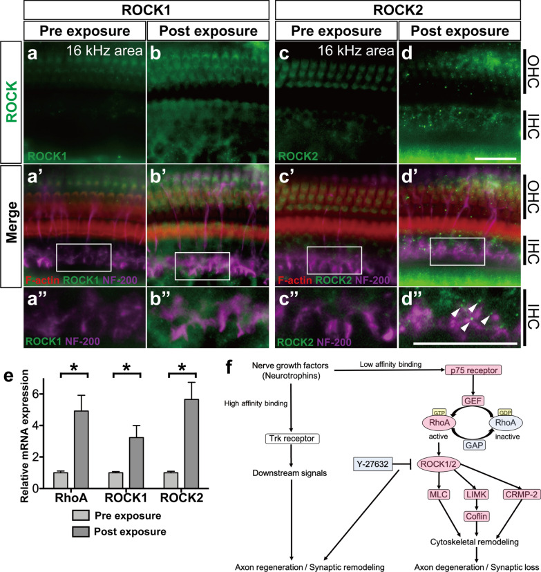
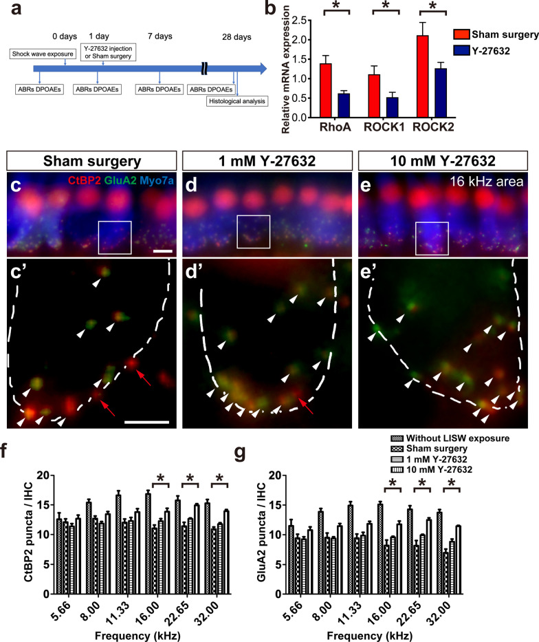
Recently, a pathological condition called cochlear synaptopathy has been clarified, and as a disorder of the auditory nerve synapses that occurs prior to failure of hair cells, it has been recognized as a major cause of sensorineural hearing loss. However, cochlear synaptopathy is untreatable. Inhibition of rho-associated coiled-coil containing protein kinase (ROCK), a serine-threonine protein kinase, has been reported to have neuroprotective and regenerative effects on synaptic pathways in the nervous system, including those in the inner ear. We previously demonstrated the regenerative effect of the ROCK inhibitor, Y-27632, on an excitotoxic cochlear nerve damage model in vitro. In this study, we aimed to validate the effect of ROCK inhibition on mice with cochlear synaptopathy induced by laser-induced shock wave (LISW) in vivo. After the elevation of ROCK1/2 expression in the damaged cochlea was confirmed, we administered Y-27632 locally via the middle ear. The amplitude of wave I in the auditory brainstem response and the number of synapses in the Y-27632-treated cochlea increased significantly. These results clearly demonstrate that ROCK inhibition has a promising clinical application in the treatment of cochlear synaptopathy, which is the major pathology of sensorineural hearing loss.

Keywords: Cochlea; Hearing loss; Inner ear; Regeneration; Rho-associated coiled-coil containing protein kinase (ROCK); Synapse; Y-27632.


Conflict of interest statement

The authors declare no competing financial interests.

Figures

Fig. 1
Fig. 1
Changes in the expression levels of ROCKs before and after cochlear damage induced by irradiation of the organ of Corti with laser-induced shock wave (LISW). (ad, a’–d’, a”–d”) Projection images of a confocal series of immunohistochemistry: tissues of the cochlear organ of Corti from the middle turn (16 kHz area) of the mouse before and 1 day after LISW exposure. Immunohistochemistry for ROCK1 (green, anti-ROCK1 (a, b), anti-ROCK2 (c, d)) and merged images with F-actin (rhodamine phalloidin, red) and neurofilament (NF-200, purple) are shown in a’-d’. (a”–d”) Enlarged images of the white inlets in (a’–d’) focusing on the inner hair cells and the end of the peripheral axons of the auditory nerve. After LISW exposure, ROCK1 expression showed ubiquitous elevation around the nerve end (b”), whereas ROCK2 expression showed punctiform enhancement around the inner hair cells (d”, white arrowheads) in addition to the ubiquitous elevation of expression. e The relative mRNA expression levels of RhoA, ROCK1, and ROCK2 before and 1 day after LISW exposure. The relative mRNA expression levels were standardized by the expression levels before LISW exposure in each mRNA (n = 6). f The schema of Rho/ROCK and associated signal pathway. *indicates a significant difference (p < 0.05, two-tailed Mann–Whitney U test). The data are shown as the mean ± standard errors of mean. CRMP-2 collapsing response mediator protein-2, GAP GTPase-activating protein, GEF guanine nucleotide-exchange factor, IHC inner hair cell, LIMK LIM kinase, MLC myosin light chain, OHC outer hair cell, Trk tropomyosin receptor kinase. Scale bar: 20 µm.
Fig. 2
Fig. 2
Effect of Y-27632 on the synapses in the inner hair cells after LISW exposure. a Timeline of the experiments in this study. b The effect of 10 mM Y-27632 in the LISW exposed cochleae on relative mRNA expression levels of RhoA, ROCK1, and ROCK2, 2 days after Y-27632 treatment. The relative mRNA expression levels were standardized by the expression levels before LISW exposure in each mRNA (n = 6). *indicates a significant difference (p < 0.05, two-tailed Mann–Whitney U test). ce The effect of Y-27632 on the degenerated synapses of the inner hair cells at cochlear 16 kHz area 28 days after treatment with various concentrations of Y-27632 (c without Y-27632, d: 1 mM, and e: 10 mM). In ce, presynaptic ribbons (CTBP2-immunoreactive puncta, red), postsynaptic densities (GluA2 immunoreactive puncta, green), and hair cells labeled with myosin 7a (blue) are shown. (c'–e') Enlarged images of the white inlets in (ce) focusing on the synapses in the inner hair cells area. White dotted lines show the contour of the inner hair cells. White arrowhead indicates normal synapse formation with CtBP2-positive patch (red) accompanied with glutamate-receptor patch (green). Red arrow indicates orphan ribbons, which is a CtBP2-positive patch without apposed glutamate-receptor patches. In c’, there are seven normal synapses (white arrowheads) and two orphan synapses (red arrows); in d’, there are 12 normal synapses and one orphan synapse; and in e’, all 14 synapses have both CtBP2 and glutamate-receptor patch. f, g The quantification of the synaptic ribbons (f) and the glutamate receptor (g) observed in the 28 days after treatment and control from single IHC. The number of synapse components is lower in the sham surgery groups than in the control group at all frequencies tested. The number of synapse components is significantly larger in the 10 mM Y-27632-treated group at higher frequencies (asterisks) than those in the sham surgery group. Scale bar is 5 µm. *indicates significant differences (p < 0.05, two-way ANOVA, followed by Bonferroni correction for multiple comparisons). Values are represented as mean ± SEM
Fig. 3
Fig. 3
Measurement of hearing function using ABR and DPOAE before and after Y-27632 treatment. ac Changes in ABR threshold at various timepoints are shown in Fig. 2a. The ABR thresholds at 28 days after Y-27632 treatment (green filled diamonds) are not significantly different from those at pre-LISW exposure (black filled circle) in any treatment groups, although the temporal threshold shifts at 1 day after LISW exposure can be observed. df ABR wave I amplitudes at the same timepoints as in a-c. Similar to the ABR threshold changes, a decrease in temporal amplitude can be observed at 1 day after LISW exposure in any groups. However, in the 1 mM Y-27632-treated group, the amplitude at 28 days after treatment (green filled diamonds) from 8 to 16 kHz is still significantly (black asterisks) lower than that at pre-LISW exposure (black filled circle). In 10 mM Y-27632 treatment group, the amplitude at 28 days after treatment (green filled diamonds) at any frequencies is not significantly different from that at pre-LISW exposure (black filled circle). However, the amplitudes at 28 days after treatment (green filled diamonds) at 8 kHz and 16 kHz are significantly larger than those at post-LISW exposure (red filled squares). gi Changes in DPOAE threshold at various timepoints are shown in Fig. 2a. No significant elevations can be observed in the DPOAE thresholds in any treatment group. j An example of 16 kHz ABR waves recorded at 28 days after Sham surgery and 10 mM Y-27632 treatment. Bilateral arrows show the wave I amplitude at 80 dB SPL. Arrowheads show the peaks with the largest peak-to-peak amplitude. In the Y-27632-treated ear, the peak could first be detected at 25 dB, while on the control side, the peak could first be detected at almost the same sound pressure level. k The representative figure of the input–output curve recorded at 28 days after Sham surgery (filled circle), 1 mM Y-27632 treatment (filled square), and 10 mM Y-27632 treatment (filled triangle). Black asterisk indicates significant changes in the value at 28 days after treatment than at pre-LISW exposure (p < 0.05, two-way ANOVA, followed by Bonferroni correction for multiple comparisons). Red asterisk indicates significant changes in the value at 28 days after treatment than at post-LISW exposure (p < 0.05, two-way ANOVA, followed by Bonferroni correction for multiple comparisons). Values are represented as mean ± SEM

References

    1. Kujawa SG, Liberman MC. Synaptopathy in the noise-exposed and aging cochlea: Primary neural degeneration in acquired sensorineural hearing loss. Hear Res. 2015;330:191–199. doi: 10.1016/j.heares.2015.02.009. - DOI - PMC - PubMed
    1. Kujawa SG, Liberman MC. Adding insult to injury: cochlear nerve degeneration after "temporary" noise-induced hearing loss. J Neurosci. 2009;29:14077–14085. doi: 10.1523/JNEUROSCI.2845-09.2009. - DOI - PMC - PubMed
    1. Sergeyenko Y, Lall K, Liberman MC, Kujawa SG. Age-related cochlear synaptopathy: an early-onset contributor to auditory functional decline. J Neurosci. 2013;33:13686–13694. doi: 10.1523/JNEUROSCI.1783-13.2013. - DOI - PMC - PubMed
    1. Dulon D, Papal S, Patni P, Cortese M, Vincent PF, Tertrais M, et al. Clarin-1 gene transfer rescues auditory synaptopathy in model of Usher syndrome. J Clin Invest. 2018;128:3382–3401. doi: 10.1172/JCI94351. - DOI - PMC - PubMed
    1. Niwa K, Mizutari K, Matsui T, Kurioka T, Matsunobu T, Kawauchi S, et al. Pathophysiology of the inner ear after blast injury caused by laser-induced shock wave. Sci Rep. 2016;6:31754. doi: 10.1038/srep31754. - DOI - PMC - PubMed

Publication types

LinkOut - more resources