

Diagnostic performance of chest radiography in high COVID-19 prevalence setting: experience from a European reference hospital
- PMID: 34218365
- PMCID: PMC8254671
- DOI: 10.1007/s10140-021-01946-x
Diagnostic performance of chest radiography in high COVID-19 prevalence setting: experience from a European reference hospital
Abstract
Purpose: The study's aim is to analyse the diagnostic performance of chest radiography (CXR) in patients with suspected coronavirus disease 19 (COVID-19).
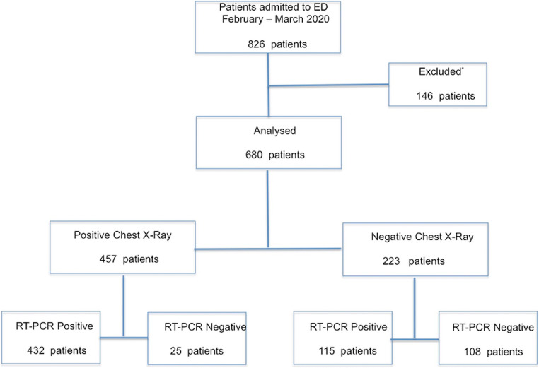
Methods: We retrospectively considered 826 consecutive patients with suspected COVID-19 presenting to our emergency department (ED) from February 21 to March 31, 2020, in a high disease prevalence setting. We enrolled patients who underwent CXR and rhino-oropharyngeal swab for real-time reverse transcription-polymerase chain reaction (rRT-PCR). CXRs were evaluated by an expert radiologist; a second independent analysis was performed by two residents in consensus. All readers, blinded to rRT-PCR results, classified CXRs positive/negative depending on presence/absence of typical findings of COVID-19, using rRT-PCR as reference standard.
Results: We finally analysed 680 patients (median age 58); 547 (80%) tested positive for COVID-19. The diagnostic performance of CXR, interpreted by the expert reader, was as follows: sensitivity (79.0%; 95% CI: 75.3-82.3), specificity (81.2%; 95% CI: 73.5-87.5), PPV (94.5%;95% CI: 92.0-96.4), NPV (48.4%; 95% CI: 41.7-55.2), and accuracy (79.3%; 95% CI: 76.0-82.2). For the residents: sensitivity (75.1%; 95% CI: 71.2-78.7), specificity (57.9%; 95% CI: 49.9-66.4), PPV (88.0%; 95% CI: 84.7-90.8), NPV (36.2%; 95% CI: 29.7-43.0), and accuracy (71.6%; 95% CI: 68.1-75.0). We found a significant difference between the reporting sensitivity (p = 0.013) and specificity (p < 0.0001) of expert radiologist vs residents. CXR sensitivity was higher in patients with symptom onset > 5 days before ED presentation compared to ≤ 5 days (84.4% vs 70.7%).
Conclusions: CXR showed a sensitivity of 79% and a specificity of 81% in diagnosing viral pneumonia in symptomatic patients with clinical suspicion of COVID-19. Further studies in lower prevalence settings are needed.
Keywords: Pneumonia; Radiology/radiography.
© 2021. The Author(s).
Conflict of interest statement
I certify that there is no conflict of interest with any financial organisation.
Figures

References
-
- Who (2021) Coronavirus disease (COVID-19) pandemic. Numbers at a glance. https://www.who.int/emergencies/diseases/novel-coronavirus-2019. Accessed 13 March 2021
-
- Bai HX, Hsieh B, Xiong Z, Halsey K, Choi JW, Tran TML, Pan I, Shi LB, Wang DC, Mei J, Jiang XL, Zeng QH, Egglin TK, Hu PF, Agarwal S, Xie FF, Li S, Healey T, Atalay MK, Liao WH. Performance of radiologists in differentiating COVID-19 from viral pneumonia on chest CT. Radiology. 2020;296:E46–E54. doi: 10.1148/radiol.2020200823. - DOI - PMC - PubMed
