. 2021 Aug 23;379(2204):20210111.
doi: 10.1098/rsta.2021.0111. Epub 2021 Jul 5.

Synergistic tomographic image reconstruction: part 2

Affiliations

Synergistic tomographic image reconstruction: part 2

Charalampos Tsoumpas et al. Philos Trans A Math Phys Eng Sci. .

Abstract

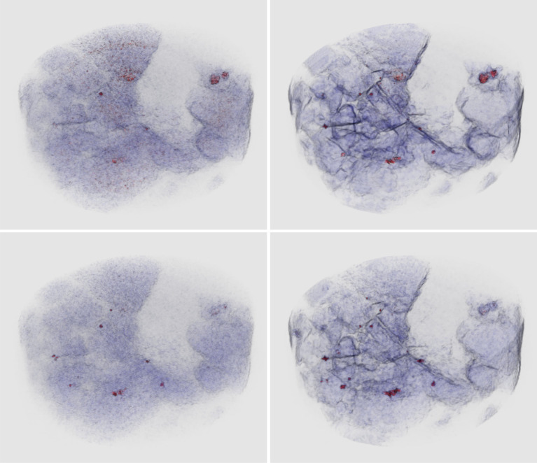
This special issue is the second part of a themed issue that focuses on synergistic tomographic image reconstruction and includes a range of contributions in multiple disciplines and application areas. The primary subject of study lies within inverse problems which are tackled with various methods including statistical and computational approaches. This volume covers algorithms and methods for a wide range of imaging techniques such as spectral X-ray computed tomography (CT), positron emission tomography combined with CT or magnetic resonance imaging, bioluminescence imaging and fluorescence-mediated imaging as well as diffuse optical tomography combined with ultrasound. Some of the articles demonstrate their utility on real-world challenges, either medical applications (e.g. motion compensation for imaging patients) or applications in material sciences (e.g. material decomposition and characterization). One of the desired outcomes of the special issues is to bring together different scientific communities which do not usually interact as they do not share the same platforms such as journals and conferences. This article is part of the theme issue 'Synergistic tomographic image reconstruction: part 2'.

Keywords: X-ray computed tomography; diffuse optical tomography; magnetic resonance imaging; open-source software; positron emission tomography; tomography.


Figures

Figure 1.
Figure 1.
Example from [5] of hyperspectral X-ray tomographic imaging in three dimensions of gold and other mineral deposits using synergistic reconstruction methods. Localizing and distinguishing inclusions with similar densities is not possible using conventional X-ray CT. New energy-sensitive X-ray detectors can, in principle, distinguish inclusions through K-edge absorption profiles which act as elemental fingerprints. However, the hyperspectral data is very noisy, causing simple reconstruction of energy channels independently to fail: channelwise SIRT reconstruction (left column) of energy channel below (top row) and above (bottom row) gold and galena K-edges are noisy and inclusions poorly defined. Using a synergistic reconstruction method described in [5] employing both spectral and spatial regularization (right column, same energy channels as before in top and bottom rows) the imaging of mineral phases and small inclusions is strongly improved. Inclusions can be distinguished due to different responses at different energies. For example, unlike the many smaller inclusions (gold and galena) the two large inclusions in the top right only show a high response at the lower energy, which helps to distinguish these as chalcopyrite. Image credit: Laura Murgatroyd, STFC, UKRI.

References

    1. Tsoumpas C, Jørgensen JS, Kolbitsch C, Thielemans K. 2021. Synergistic tomographic image reconstruction: part 1. Phil. Trans. R. Soc. A 379, 20200189. (10.1098/rsta.2020.0189) - DOI - PMC - PubMed
    1. Arridge SR, Ehrhardt MJ, Thielemans K. 2021. (An overview of) Synergistic reconstruction for multimodality/multichannel imaging methods. Phil. Trans. R. Soc. A 379, 20200205. (10.1098/rsta.2020.0205) - DOI - PubMed
    1. Polycarpou I, Soultanidis G, Tsoumpas C. 2021. Synergistic motion compensation strategies for positron emission tomography when acquired simultaneously with magnetic resonance imaging. Phil. Trans. R. Soc. A 379, 20200207. (10.1098/rsta.2020.0207) - DOI - PMC - PubMed
    1. Jørgensen JS et al. 2021. Core Imaging Library part I: a versatile Python framework for tomographic imaging. Phil. Trans. R. Soc. A 379, 20200192. (10.1098/rsta.2020.0192) - DOI - PMC - PubMed
    1. Papoutsellis E et al. 2021. Core Imaging Library – Part II: multichannel reconstruction for dynamic and spectral tomography. Phil. Trans. R. Soc. A 379, 20200193. (10.1098/rsta.2020.0193) - DOI - PMC - PubMed

Publication types

MeSH terms

LinkOut - more resources