. 2021 Aug 23;379(2204):20200192.
doi: 10.1098/rsta.2020.0192. Epub 2021 Jul 5.

Core Imaging Library - Part I: a versatile Python framework for tomographic imaging

Affiliations

Core Imaging Library - Part I: a versatile Python framework for tomographic imaging

J S Jørgensen et al. Philos Trans A Math Phys Eng Sci. .

Abstract

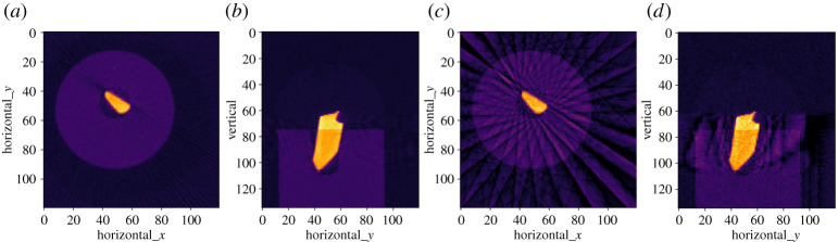
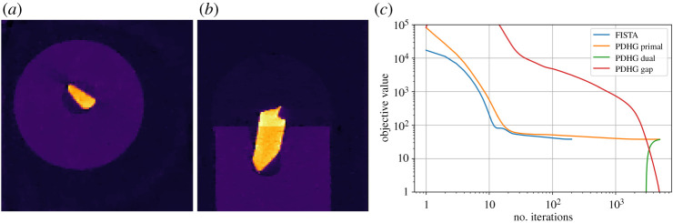
We present the Core Imaging Library (CIL), an open-source Python framework for tomographic imaging with particular emphasis on reconstruction of challenging datasets. Conventional filtered back-projection reconstruction tends to be insufficient for highly noisy, incomplete, non-standard or multi-channel data arising for example in dynamic, spectral and in situ tomography. CIL provides an extensive modular optimization framework for prototyping reconstruction methods including sparsity and total variation regularization, as well as tools for loading, preprocessing and visualizing tomographic data. The capabilities of CIL are demonstrated on a synchrotron example dataset and three challenging cases spanning golden-ratio neutron tomography, cone-beam X-ray laminography and positron emission tomography. This article is part of the theme issue 'Synergistic tomographic image reconstruction: part 2'.

Keywords: X-ray CT; computed tomography; convex optimization; image reconstruction; software.


Figures

Figure 1.
Figure 1.
Overview of CIL module structure and contents. The cil.plugins module contains wrapper code for other software and third-party libraries that need to be installed separately to be used by CIL.
Figure 2.
Figure 2.
Raw and preprocessed three-dimensional parallel-beam X-ray CT steel-wire dataset. (a) Raw transmission projection. (b) Scaled, cropped, centred and negative-log transformed projection. (c)(i) Sinogram for slice vertical=103, all 90 angles. (c)(ii) Same, subsampled to 15 equi-spaced angles.
Figure 9.
Figure 9.
CIL AcquisitionGeometry and ImageGeometry illustrated for the laminography cone-beam setup. Configurable parameters are shown in the legend. Parallel-beam geometry and two-dimensional versions are also available. CIL can illustrate ImageGeometry and AcquisitionGeometry instances as in this figure using show_geometry(ag,ig).
Figure 3.
Figure 3.
Reconstructions of steel-wire dataset by FBP. (a,b) Horizontal and vertical slices using 90 projections. (c,d) Same using 15 projections—showing prominent streak artefacts. Colour range [−0.01, 0.11].
Figure 4.
Figure 4.
Algebraic iterative reconstruction of 15-projection three-dimensional steel-wire dataset. (a,b) Horizontal and vertical slices, 20-iteration CGLS reconstruction. (c,d) Same using SIRT, lower/upper bounds 0.0/0.09. Colour range [−0.01, 0.11].
Figure 5.
Figure 5.
Anisotropic Tikhonov reconstruction of 15-projection three-dimensional steel-wire dataset. (a,b) Horizontal and vertical slices, Tikhonov regularization with horizontal smoothing (αx = αy = 30, αz = 0.1). (c,d) Same, with vertical smoothing (αx = αy = 0.1, αz = 60). Colour range [−0.01, 0.11].
Figure 6.
Figure 6.
FISTA reconstruction of 15-projection three-dimensional steel-wire dataset. (a,b) L1-norm regularization with large regularization parameter of α = 30 forces all pixels but in steel wire to zero. (c,d) TV-regularization with α = 0.02 removes streaks and noise and preserves edges. Colour range [−0.01, 0.11].
Figure 7.
Figure 7.
PDHG reconstruction of 15-projection three-dimensional steel-wire dataset. (a,b) TV-regularization with α = 0.02, reproduces the same result as FISTA in figure 6 on the same case and parameter choice, thus validating algorithms against each other. Colour range [−0.01, 0.11]. (c) Objective value histories (log-log) for FISTA and PDHG on TV-regularization problem. Both algorithms reach the same (primal) objective value, FISTA taking fewer but slower iterations. The primal-dual gap for PDHG (difference between primal and dual objectives) approaches zero indicating convergence.
Figure 8.
Figure 8.
IMAT neutron tomography dataset. Top row: (left) top-view schematic of high-purity elemental metal rod sample; (centre) top-view photograph; (right) single raw projection image showing rods of different absorption. Middle row: (left) preprocessed slice sinogram; (right) horizontal line profile of FBP, PDHG TV and GD TV reconstruction along line shown on image below. Bottom row: (left) slice reconstructions, FBP; (centre) TV reconstruction with PDHG; (right) STV reconstruction with GD. Colour range [−0.002, 0.012].
Figure 10.
Figure 10.
Slices through three-dimensional reconstruction of laminography LEGO sample. Left, top/bottom: LS reconstruction using FISTA, horizontal/vertical slice at yellow line. Right: Same using TVNN, in which laminography artefacts are suppressed while edges are preserved.
Figure 11.
Figure 11.
Three-dimensional PET reconstruction of NEMA IQ phantom data using CIL with SIRF data structures. (a) OSEM reconstruction (SIRF), horizontal slice. (b) KLTV reconstruction (CIL PDHG). Colour range both [0,0.15]. (c) OSEM and KLTV profiles along red vertical line on centre plot.

References

    1. Shearing P, Turner M, Sinclair I, Lee P, Ahmed F, Quinn P, Leach R, Sun W, Warnett J. 2018. EPSRC X-Ray Tomography Roadmap 2018. Available from: https://epsrc.ukri.org/files/research/epsrc-x-ray-tomography-roadmap-2018/.
    1. Pan X, Sidky EY, Vannier M. 2009. Why do commercial CT scanners still employ traditional, filtered back-projection for image reconstruction? Inverse Prob. 25, 123009. (10.1088/0266-5611/25/12/123009) - DOI - PMC - PubMed
    1. Zhang H, Wang J, Zeng D, Tao X, Ma J. 2018. Regularization strategies in statistical image reconstruction of low-dose x-ray CT: a review. Med. Phys. 45, e886-e907. (10.1002/mp.13123) - DOI - PMC - PubMed
    1. Ametova E, Fardell G, Jørgensen JS, Papoutsellis E, Pasca E. 2021. Releases of Core Imaging Library (CIL). Zenodo. (10.5281/zenodo.4746198) - DOI
    1. Gürsoy D, De Carlo F, Xiao X, Jacobsen C. 2014. TomoPy: a framework for the analysis of synchrotron tomographic data. J. Synchrotron Radiat. 21, 1188-1193. (10.1107/S1600577514013939) - DOI - PMC - PubMed

MeSH terms