. 2021 Jun 18:15:680549.
doi: 10.3389/fncom.2021.680549. eCollection 2021.

Computational Evidence for a Competitive Thalamocortical Model of Spikes and Spindle Activity in Rolandic Epilepsy

Affiliations

Computational Evidence for a Competitive Thalamocortical Model of Spikes and Spindle Activity in Rolandic Epilepsy

Qiang Li et al. Front Comput Neurosci. .

Abstract

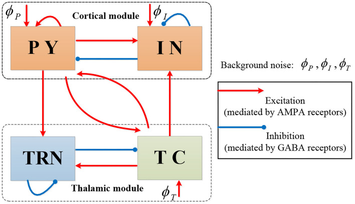
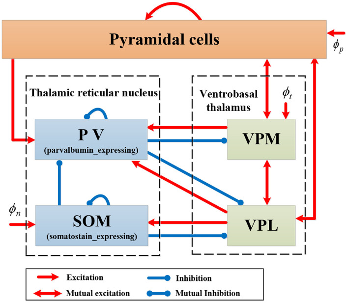
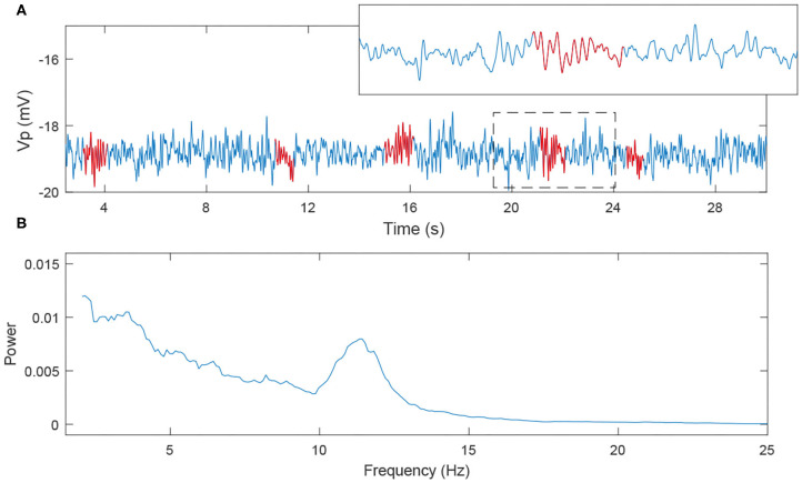
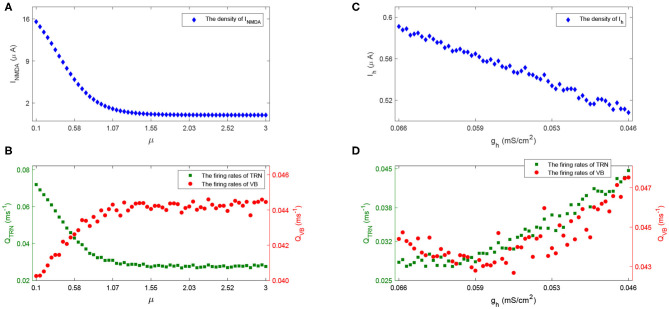
Rolandic epilepsy (RE) is the most common idiopathic focal childhood epilepsy syndrome, characterized by sleep-activated epileptiform spikes and seizures and cognitive deficits in school age children. Recent evidence suggests that this disease may be caused by disruptions to the Rolandic thalamocortical circuit, resulting in both an abundance of epileptiform spikes and a paucity of sleep spindles in the Rolandic cortex during non-rapid eye movement sleep (NREM); electrographic features linked to seizures and cognitive symptoms, respectively. The neuronal mechanisms that support the competitive shared thalamocortical circuitry between pathological epileptiform spikes and physiological sleep spindles are not well-understood. In this study we introduce a computational thalamocortical model for the sleep-activated epileptiform spikes observed in RE. The cellular and neuronal circuits of this model incorporate recent experimental observations in RE, and replicate the electrophysiological features of RE. Using this model, we demonstrate that: (1) epileptiform spikes can be triggered and promoted by either a reduced NMDA current or h-type current; and (2) changes in inhibitory transmission in the thalamic reticular nucleus mediates an antagonistic dynamic between epileptiform spikes and spindles. This work provides the first computational model that both recapitulates electrophysiological features and provides a mechanistic explanation for the thalamocortical switch between the pathological and physiological electrophysiological rhythms observed during NREM sleep in this common epileptic encephalopathy.

Keywords: BECTS; CECTS; Costa neural mass model; benign epilepsy with centrotemporal spikes; childhood epilepsy; electroencephalogram; neural mass model.


Conflict of interest statement

The authors declare that the research was conducted in the absence of any commercial or financial relationships that could be construed as a potential conflict of interest.

Figures

Figure 1
Figure 1
The topological structure of Costa model.
Figure 2
Figure 2
The dynamical evolution in each population in Costa model.
Figure 3
Figure 3
Schematic representation of model RE-NMM.
Figure 4
Figure 4
Example data analysis pipeline. (A) NREM sleep data segment from one clinical EEG. The red circles represent detected spikes. (B) The power spectra computed from a 60 s epoch. We note that in this epoch where spikes are abundant, the expected sigma bump reflecting sleep spindles is absent. (C) The histogram of inter-spike intervals computed from the same data epoch.
Figure 5
Figure 5
The simulated EEGs (i.e., the model output Vp) as well as the histogram of inter-spike intervals under different situations. (A) μ = 0.5 and gh = 0.066; (B) μ = 2 and gh = 0.066; (C) μ = 0.5 and gh = 0.056; (D) μ = 2 and gh = 0.056.
Figure 6
Figure 6
The evolution of spike rates in the parameter space μ × gh.
Figure 7
Figure 7
The simulated EEG signal and its power spectra. (A) 30s EEG segment; (B) power spectra (the red labeled part corresponds to the spindles).
Figure 8
Figure 8
The evolution of spike rate (A) and spindle rate (B) in parameter space Nt1r1 × Nt2r1 × Nt2r2 ∈ [1, 5.5] × [1, 2] × [1, 2].
Figure 9
Figure 9
(A) The combinations of spike rate and spindle rate in the diagonal grids of the parameter space Nt1r1 × Nt2r1 × Nt2r2 ∈ [1, 5.5] × [1, 2] × [1, 2]. (B) 60s simulated EEG segment with high spindle rate and low spike rate (corresponding to the point A); (C) 60s simulated EEG segment with high spike rate and low spindle rate (corresponding to the point B) (the red labeled part represents the spindles, the green circle represents the spikes).
Figure 10
Figure 10
(A) PV cells (top) have increased firing compared to SOM cells (bottom). Boxplots of Kruskal-Wallis test statistic for the spindle rate (B) and spike rate (C) with respect to three cases (PV+SOM, only PV, only SOM).
Figure 11
Figure 11
The spike rate distribution of 27 real RE-EEG segments in the estimated parameter space μ^×ĝh.
Figure 12
Figure 12
An example of the model fit to one real RE-EEG segment. (A) A 60s real RE-EEG segment from “patient 1”; (B) The simulated 60s EEG segment; (C,D) Comparison between the features extracted from the real EEG and the simulated one.
Figure 13
Figure 13
The density of INMDA (A) and firing rate of population TRN and VB (B) with the increase of μ; The density of Ih (C) and firing rate of TRN and VB (D) with the decrease of gh.

References

    1. Ahrens S., Jaramillo S., Yu K., Ghosh S., Hwang G., Paik R., et al. . (2015). ErbB4 regulation of a thalamic reticular nucleus circuit for sensory selection. Nat. Neurosci. 18, 104–111. 10.1038/nn.3897 - DOI - PMC - PubMed
    1. Archer J. S., Briellman R. S., Abbott D. F., Syngeniotis A., Wellard R. M., Jackson G. D. (2003). Benign epilepsy with centro-temporal spikes: spike triggered fMRI shows somato-sensory cortex activity. Epilepsia 44, 200–204. 10.1046/j.1528-1157.2003.02502.x - DOI - PubMed
    1. Babadi B., Brown E. N. (2014). A review of multitaper spectral analysis. IEEE Trans. Biomed. Eng. 61, 1555–1564. 10.1109/TBME.2014.2311996 - DOI - PubMed
    1. Beenhakker M. P., Huguenard J. R. (2009). Neurons that fire together also conspire together: is normal sleep circuitry hijacked to generate epilepsy? Neuron 62, 612–632. 10.1016/j.neuron.2009.05.015 - DOI - PMC - PubMed
    1. Brennan G. P., Baram T. Z., Poolos N. P. (2016). Hyperpolarization-activated cyclic nucleotide-gated (HCN) channels in epilepsy. Cold Spring Harbor Perspect. Med. 6:a022384. 10.1101/cshperspect.a022384 - DOI - PMC - PubMed

LinkOut - more resources