. 2021 Jun 17:12:653232.
doi: 10.3389/fphar.2021.653232. eCollection 2021.

Cryptotanshinone Inhibits the Growth of HCT116 Colorectal Cancer Cells Through Endoplasmic Reticulum Stress-Mediated Autophagy

Affiliations

Cryptotanshinone Inhibits the Growth of HCT116 Colorectal Cancer Cells Through Endoplasmic Reticulum Stress-Mediated Autophagy

Xiaojing Fu et al. Front Pharmacol. .

Abstract

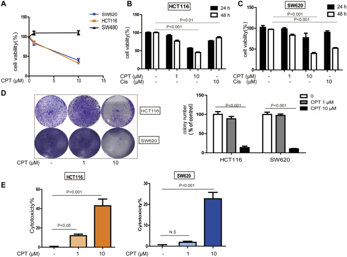
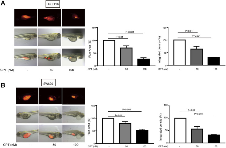
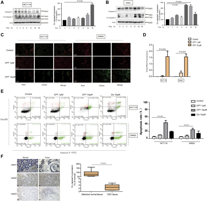
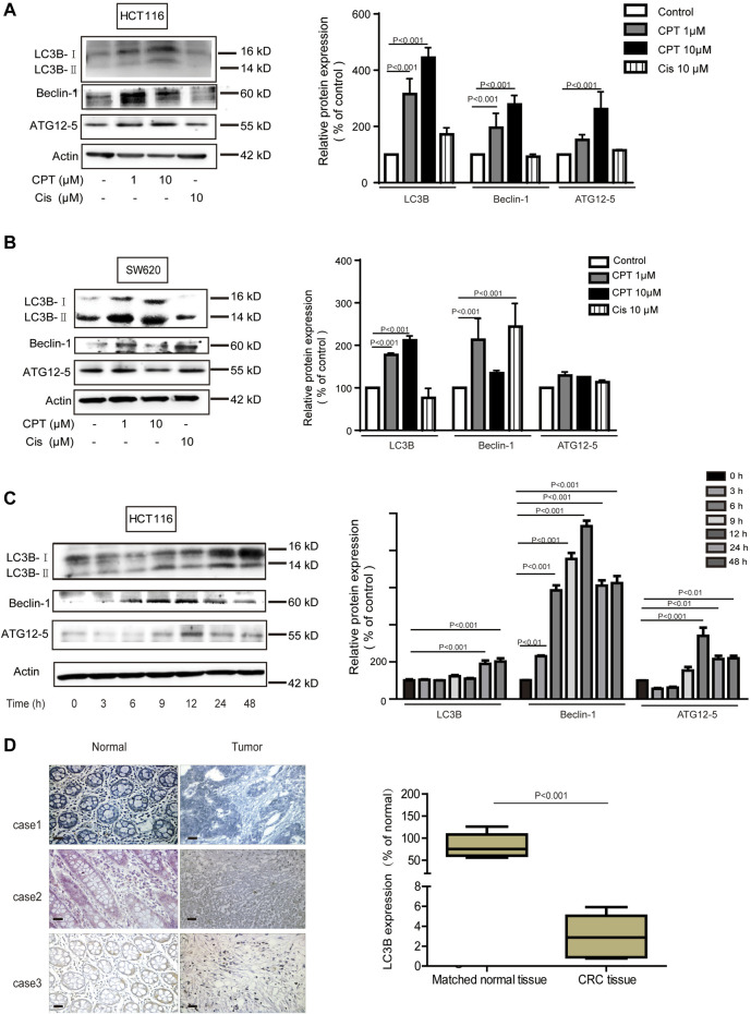
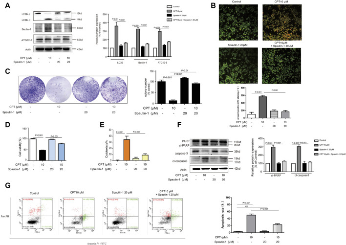
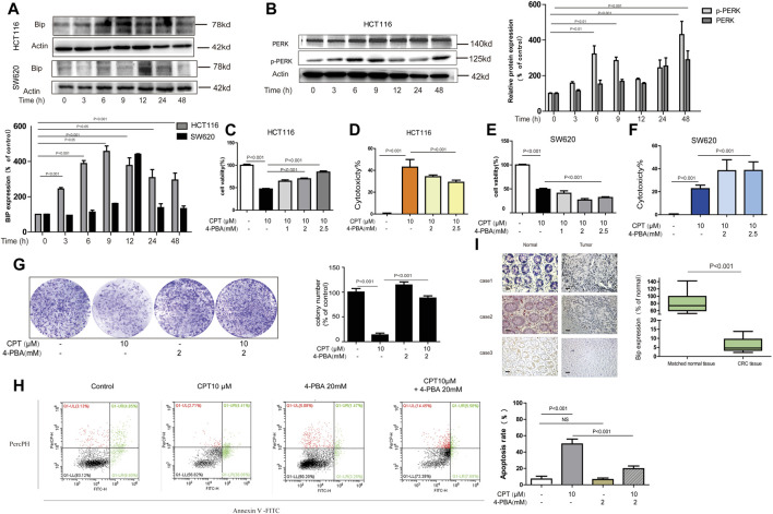
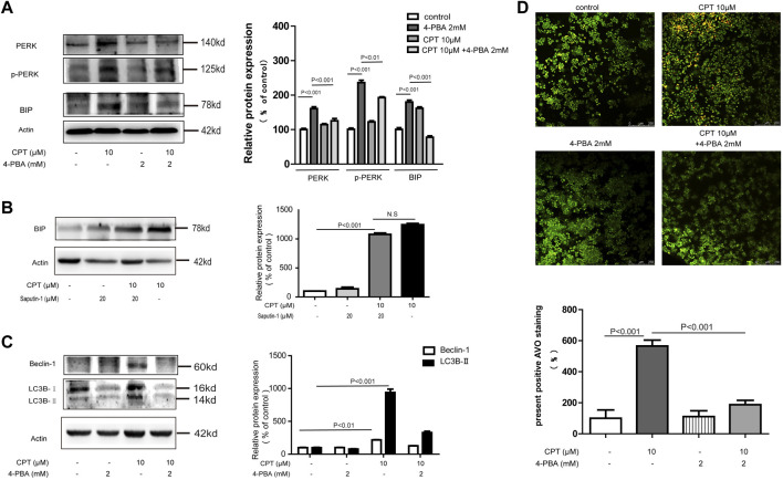
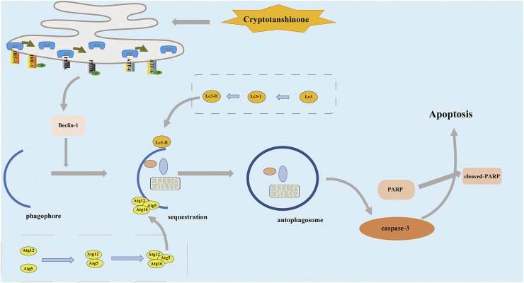
Among cancers, colorectal cancer (CRC) has one of the highest annual incidence and death rates. Considering severe adverse reactions associated with classical chemotherapy medications, traditional Chinese medicines have become potential drug candidates. In the current study, the effects of cryptotanshinone (CPT), a major component of Salvia miltiorrhiza Bunge (Danshen) on CRC and underlying mechanism were explored. First of all, data from in vitro experiments and in vivo zebrafish models indicated that CPT selectively inhibited the growth and proliferation of HCT116 and SW620 cells while had little effect on SW480 cells. Secondly, both ER stress and autophagy were associated with CRC viability regulation. Interestingly, ER stress inhibitor and autophagy inhibitor merely alleviated cytotoxic effects on HCT116 cells in response to CPT stimulation, while have little effect on SW620 cells. The significance of apoptosis, autophagy and ER stress were verified by clinical data from CRC patients. In summary, the current study has revealed the anti-cancer effects of CPT in CRC by activating autophagy signaling mediated by ER stress. CPT is a promising drug candidate for CRC treatment.

Keywords: apoptosis; autophagy; colorectal cancer; cryptotanshinone; endoplasmic reticulum stress.


Conflict of interest statement

The authors declare that the research was conducted in the absence of any commercial or financial relationships that could be construed as a potential conflict of interest.

Figures

FIGURE 1
FIGURE 1
The role of CPT against colorectal cancer. MTT assay (A) was used to measure the activity of SW480, HCT116, and SW620 cells treated with concentrations of CPT for 48 h. SW620 and HCT116 were cultured in different concentrations of CPT or cisplatin (Cis) for 24 or 48 h, respectively, and cell proliferation rates were analyzed by MTT assay (B,C). Colony formation assay of SW620 and HCT116 cells treated with the indicated concentrations of CPT. Representative images (Left) and quantification of colonies (Right) were shown (D). HCT116 cells and SW620 cells were treated with different concentrations of CPT for 48 h, and the release amount of lactate dehydrogenase was analyzed by LDH assay, and the cytotoxicity was calculated (E,F). CPT, cryptotanshinone; LDH, lactate dehydrogenase; Cis, cisplatin.
FIGURE 2
FIGURE 2
The inhibitory effects of different concentrations of CPT on tumor growth of HCT116 and SW620 zebrafish models were determined. The inhibitory effects of different concentrations of CPT on tumor growth of HCT116 and SW620 zebrafish were determined. The tumor was indicated by red fluorescence (A), calculated as a percentage of the control value (n = 8) (B). CPT, cryptotanshinone.
FIGURE 3
FIGURE 3
CPT can induce apoptosis of CRC cells. Caspase 3 and cleaved caspase3 in the lysates of HCT116 and SW620 cells treated with CPT (10 μM) for different time were detected by western blot assay and quantified by Image J (A,B). The JC-1 assay was used to detect HCT116 cells and SW620 cells treated with different concentrations of CPT for 48 h. The change of red or green color was observed under fluorescence microscope (200 ×) (C) and green/red was calculated (D) (n = 3). Apoptosis of HCT116 cells and SW620 cells treated as in (C) was determined by Annexin Ⅴ-FITC/PI staining (E). The immunohistochemical analysis of the clinical samples were performed, and the expression of cleaved caspase3 was detected in the tumor tissue and the corresponding para-cancer tissue, and the quantification analysis was performed with Image J (F). CPT, cryptotanshinone; CRC, colorectal cancer.
FIGURE 4
FIGURE 4
In CRC cells, autophagy mediates the anti-tumor effect of CPT. LC3B, Beclin-1, and ATG12–5 of HCT116 cells (A) and SW620 cells (B) treated with different concentrations of CPT or Cis for 48 h was detected by immunoblotting and quantitatively analyzed by ImageJ. LC3B, Beclin-1, and ATG12–5 in HCT116 cells treated with CPT at a concentration of 10 μM for different times was detected by western blot and quantitatively analyzed by Image J (C). The immunohistochemical analysis of the clinical samples were performed, and the expression of LC3 was detected in the tumor tissue and the corresponding para-cancer tissue, and the quantification analysis was performed with Image J (D). CPT, cryptotanshinone; CRC, colorectal cancer.
FIGURE 5
FIGURE 5
CPT can induce autophagy - mediated apoptosis in CRC cells. HCT116 cells were treated with or without CPT (10 μM) for 48 h in culture medium in the presence or absence of spautin-1 (20 μM). The expression of autophagy-related proteins (ATG12–5, Beclin-1, LC3B) in cell lysate was measured by western blot (A). Fluorescence microscopy (200 ×) following AO staining detected the number of autophagosomes in HCT116 cells treated as in (A), and quantitative analysis was performed by ImageJ (B). In the presence or absence of spautin-1 (20 μM), the colony formation assay of HCT116 cells treated with or without 10 μM CPT was performed, and the left image was representative and the right image was quantitative (C). MTT assay (D), and LDH release (E) assay were used to determine HCT116 cells treated as in (C). Caspase-3 and PARP in HCT116 cells treated as in (A) were analyzed by western blotting assay and quantitatively analyzed by ImageJ (F). Apoptosis of HCT116 cells treated as in (C) was quantified by flow cytometry (G). CPT, cryptotanshinone; LDH, lactate dehydrogenase; CRC, colorectal cancer; AO, acridine orang.
FIGURE 6
FIGURE 6
In HCT116 cell, ER stress mediates the anti-tumor effect of CPT. Western blot was used to determine BIP in HCT116 cells and SW620 cell lysates treated with CPT (10 μM) for different times (A). PERK and p-PERK was detected in HCT116 cells treated as in (A) using western blotting analysis (B). In the presence or absence of 4-PBA (1, 2, and 2.5 mM), HCT116 cells, and SW620 cells treated or untreated with CPT (10 μM) were cultured for 48 h and tested by MTT assay (C, E) and LDH assay (D, F). In the presence or absence of 4-PBA (2 mM), the colony formation assay of HCT116 cells treated with or without CPT (10 μM) was performed and quantified with Image J (G). Apoptosis of HCT116 cells treated as in (G) was detected by flow cytometry (H). The immunohistochemical analysis of the clinical samples were performed, and the expression of BIP was detected in the tumor tissue and the corresponding para-cancer tissue, and the quantification analysis was performed with Image J (I). CPT, cryptotanshinone; ER stress, endoplasmic reticulum stress; LDH, lactate dehydrogenase.
FIGURE 7
FIGURE 7
CPT induced ER stress mediated autophagy in HCT116 cells. Expression levels of PERK and P-PERK in HCT116 cells treated with or without 4-PBA (2 mM) in the presence or absence of CPT (10 μM) (A). HCT116 cells treated with or without Spautin-1 (20 μM) for 48 h were treated with or without CPT (10 μM). The expression of BIP in the lysate was determined by western blot and quantified by Image J (B). To test for Beclin-1 and LC3 expression in 10 μM CPT processed or untreated HCT116 cells, with or without 4-PBA (2 mM) (C). Under fluorescence microscope (200 ×), the number of autophagosomes in HCT116 cells was stained by AO staining and quantified by ImageJ software (D). CPT, cryptotanshinone; ER stress, endoplasmic reticulum stress; AO, acridine orang.
FIGURE 8
FIGURE 8
In the treatment of HCT116 cells by CPT, the apoptosis pathway is activated, which is associated with endoplasmic reticulum stress, and autophagy. CPT, cryptotanshinone.

Similar articles

Cited by

References

    1. Alper P., Salomatina O. V., Salakhutdinov N. F., Ulukaya E., Ari F. (2021). Soloxolone Methyl, as a 18βH-Glycyrrhetinic Acid Derivate, May Result in Endoplasmic Reticulum Stress to Induce Apoptosis in Breast Cancer Cells. Bioorg. Med. Chem. 30, 115963. 10.1016/j.bmc.2020.115963 - DOI - PubMed
    1. Bhardwaj M., Leli N. M., Koumenis C., Amaravadi R. K. (2019). Regulation of Autophagy by Canonical and Non-canonical ER Stress Responses. Semin. Cancer Biol. 66, 116-128. 10.1016/j.semcancer.2019.11.007 - DOI - PMC - PubMed
    1. Cai Y., Zhang W., Chen Z., Shi Z., He C., Chen M. (2016). Recent Insights into the Biological Activities and Drug Delivery Systems of Tanshinones. Int. J. Nanomedicine 11, 121–130. 10.2147/ijn.S84035 - DOI - PMC - PubMed
    1. Chen L., Yang Q., Zhang H., Wan L., Xin B., Cao Y., et al. (2020). Cryptotanshinone Prevents Muscle Wasting in CT26-induced Cancer Cachexia through Inhibiting STAT3 Signaling Pathway. J. Ethnopharmacol 260, 113066. 10.1016/j.jep.2020.113066 - DOI - PubMed
    1. Chen X., Guo J., Bao J., Lu J., Wang Y. (2014). The Anticancer Properties of Salvia Miltiorrhiza Bunge (Danshen): a Systematic Review. Med. Res. Rev. 34 (4), 768–794. 10.1002/med.21304 - DOI - PubMed

LinkOut - more resources