. 2021 Jun 18:12:679610.
doi: 10.3389/fphar.2021.679610. eCollection 2021.

Light-Responsive Micelles Loaded With Doxorubicin for Osteosarcoma Suppression

Affiliations

Light-Responsive Micelles Loaded With Doxorubicin for Osteosarcoma Suppression

Jiayi Chen et al. Front Pharmacol. .

Abstract

The enhancement of tumor targeting and cellular uptake of drugs are significant factors in maximizing anticancer therapy and minimizing the side effects of chemotherapeutic drugs. A key challenge remains to explore stimulus-responsive polymeric nanoparticles to achieve efficient drug delivery. In this study, doxorubicin conjugated polymer (Poly-Dox) with light-responsiveness was synthesized, which can self-assemble to form polymeric micelles (Poly-Dox-M) in water. As an inert structure, the polyethylene glycol (PEG) can shield the adsorption of protein and avoid becoming a protein crown in the blood circulation, improving the tumor targeting of drugs and reducing the cardiotoxicity of doxorubicin (Dox). Besides, after ultraviolet irradiation, the amide bond connecting Dox with PEG can be broken, which induced the responsive detachment of PEG and enhanced cellular uptake of Dox. Notably, the results of immunohistochemistry in vivo showed that Poly-Dox-M had no significant damage to normal organs. Meanwhile, they showed efficient tumor-suppressive effects. This nano-delivery system with the light-responsive feature might hold great promises for the targeted therapy for osteosarcoma.

Keywords: doxorubicin; light-responsive nanoparticles; micelles; osteosarcoma; targeted therapy.


Conflict of interest statement

The authors declare that the research was conducted in the absence of any commercial or financial relationships that could be construed as a potential conflict of interest.

Figures

SCHEME 1
SCHEME 1
Schematic illustration of the self-assembly and responsive breakage of Poly-Dox micelles, and the process of tumor therapy.
FIGURE 1
FIGURE 1
(A) Detailed depolymerization routes of Poly-Dox copolymer drug conjugates. (B) 1H-NMR spectra of Poly-Dox in DMSO-d6. (C) TEM image of Poly-Dox-M. (D) TEM image of disassembled Poly-Dox-M after UV irradiation. (E) Fluorescence intensity of Free-Dox, Poly-Dox-M, and Poly-Dox-M/UV. (F) The fluorescence intensity changes of Poly-Dox-M after UV irradiation of 0, 3, 5, 7, 10, 13, 16, and 20 min.
FIGURE 2
FIGURE 2
(A) K7M2-wt cells stained by Hoechst 33,342 after treated with Poly-Dox-M for 2, 4, and 6 h under UV irradiation or not. Blue signal reflects nucleus; red signal reflects Dox. Scale bar, 50 μm. (B) Their fluorescence intensity of Poly-Dox-M in different groups (Student’s t-test, *p < 0.05, **p < 0.01, ***p < 0.001).
FIGURE 3
FIGURE 3
Cell viability of K7M2-wt cells treated with (A) Free-Dox. (B) Poly-Dox-M and (C) Poly-Dox-M under UV irradiation at a various dose of Dox or Poly-Dox. (D) K7M2-wt cells stained by calcein-AM (green) and PI (red) after 6 h incubation with PBS, free Dox, Poly-Dox-M under UV irradiation or not. Blue signal represents live cell, red signal represents dead cell. Scale bar: 50 μm.
FIGURE 4
FIGURE 4
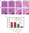
(A) H&E staining of tumor sections extracted from the mice treated with different samples. Scale bar: 20 μm. (B) An enlarged view of the boxed region below the corresponding image. Scale bar, 20 μm. (C) Image J analysis of H&E staining (Student’s t-test, *p < 0.05, **p < 0.01, ***p < 0.001).
FIGURE 5
FIGURE 5
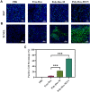
Immunofluorescent staining of tumor sections extracted from the mice treated with different samples. (A) Images of immunofluorescent staining for Ki67. Blue signals represent nucleus, green signals represent ki67 expression. (B) Images of Tunel staining. Blue signals represent nucleus, green signals represent apoptotic cells. (C) ImageJ analysis of TUNEL positive cell percentage. (Student’s t-test, *p < 0.05, **p < 0.01, ***p < 0.001).
FIGURE 6
FIGURE 6
H&E staining of various tissues including heart, liver, spleen, lung and kidney extracted from the mice treated with PBS, Free-Dox, Poly-Dox-M under UV irradiation or not.

Similar articles

Cited by

References

    1. Alvarez-Lorenzo C., Bromberg L., Concheiro A. (2009). Light-sensitive Intelligent Drug Delivery Systems. Photochem. Photobiol. 85 (4), 848–860. 10.1111/j.1751-1097.2008.00530.x - DOI - PubMed
    1. Andreadis C., Gimotty P. A., Wahl P., Hammond R., Houldsworth J., Schuster S. J., et al. (2007). Members of the Glutathione and ABC-Transporter Families Are Associated with Clinical Outcome in Patients with Diffuse Large B-Cell Lymphoma. Blood 109 (8), 3409–3416. 10.1182/blood-2006-09-047621 - DOI - PMC - PubMed
    1. Anninga J. K., Gelderblom H., Fiocco M., Kroep J. R., Taminiau A. H., Hogendoorn P. C., et al. (2011). Chemotherapeutic Adjuvant Treatment for Osteosarcoma: where Do We Stand?. Eur. J. Cancer 47 (16), 2431–2445. 10.1016/j.ejca.2011.05.030 - DOI - PubMed
    1. Beaute L., McClenaghan N., Lecommandoux S. (2019). Photo-triggered Polymer Nanomedicines: From Molecular Mechanisms to Therapeutic Applications. Adv. Drug Deliv. Rev. 138, 148–166. 10.1016/j.addr.2018.12.010 - DOI - PubMed
    1. Berrino F., De Angelis R., Sant M., Rosso S., Bielska-Lasota M., Coebergh J. W., et al. (2007). Survival for Eight Major Cancers and All Cancers Combined for European Adults Diagnosed in 1995-99: Results of the EUROCARE-4 Study. Lancet Oncol. 8 (9), 773–783. 10.1016/S1470-2045(07)70245-0 - DOI - PubMed