

Differences Changes in Cerebellar Functional Connectivity Between Mild Cognitive Impairment and Alzheimer's Disease: A Seed-Based Approach
- PMID: 34220669
- PMCID: PMC8248670
- DOI: 10.3389/fneur.2021.645171
Differences Changes in Cerebellar Functional Connectivity Between Mild Cognitive Impairment and Alzheimer's Disease: A Seed-Based Approach
Abstract
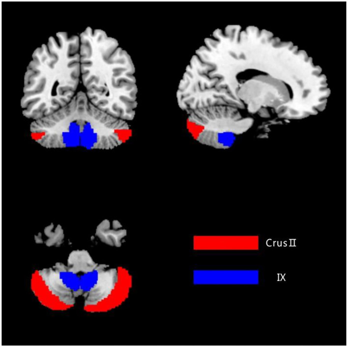
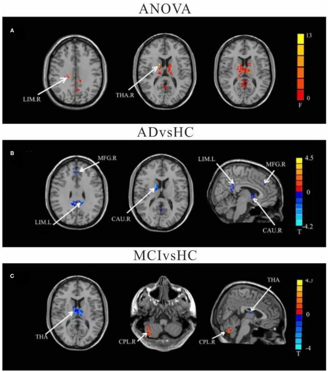
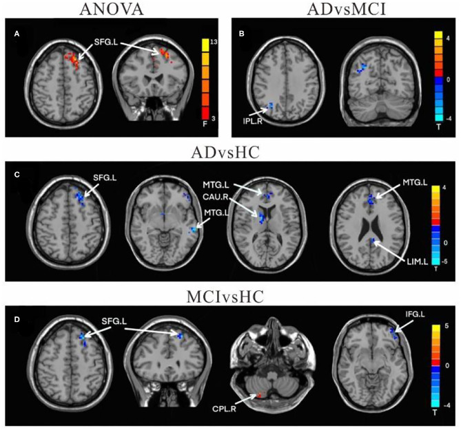
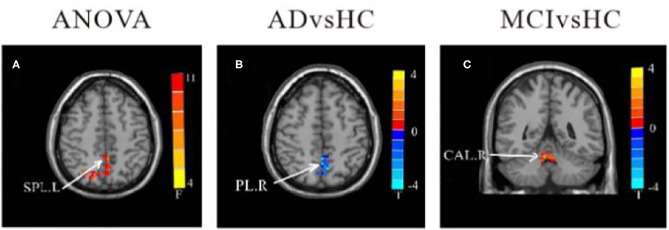
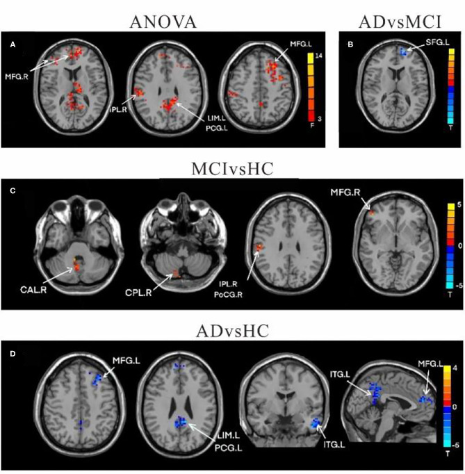
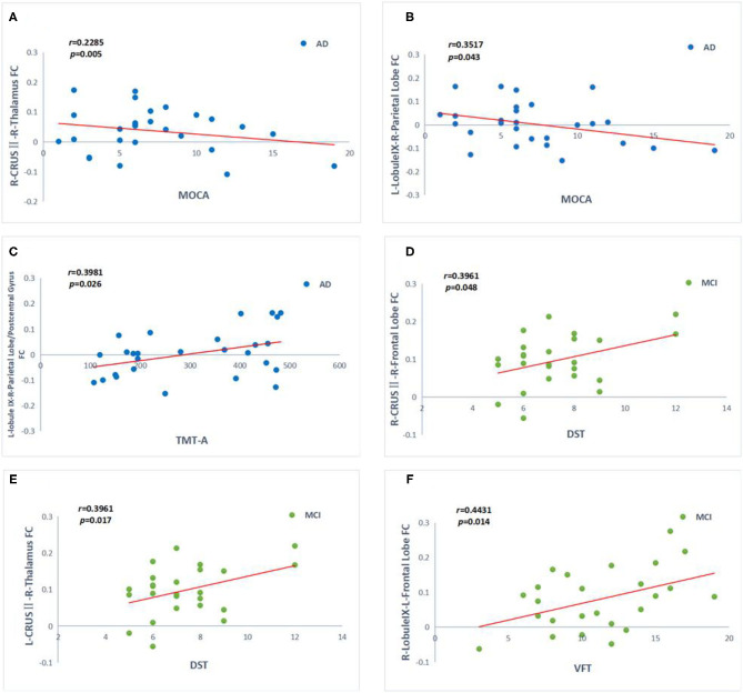
Background: Recent studies have discovered that functional connections are impaired among patients with Alzheimer's disease (AD), even at the preclinical stage. The cerebellum has been implicated as playing a role in cognitive processes. However, functional connectivity (FC) among cognitive sub-regions of the cerebellum in patients with AD and mild cognitive impairment (MCI) remains to be further elucidated. Objective: Our study aims to investigate the FC changes of the cerebellum among patients with AD and MCI, compared to healthy controls (HC). Additionally, we explored the role of cerebellum FC changes in the cognitive performance of all subjects. Materials: Resting-state functional magnetic resonance imaging (rs-fMRI) data from three different groups (28 AD patients, 26 MCI patients, and 30 HC) was collected. We defined cerebellar crus II and lobule IX as seed regions to assess the intragroup differences of cortico-cerebellar connectivity. Bias correlational analysis was performed to investigate the relationship between changes in FC and neuropsychological performance. Results: Compared to HC, AD patients had decreased FC within the caudate, limbic lobe, medial frontal gyrus (MFG), middle temporal gyrus, superior frontal gyrus, parietal lobe/precuneus, inferior temporal gyrus, and posterior cingulate gyrus. Interestingly, MCI patients demonstrated increased FC within inferior parietal lobe, and MFG, while they had decreased FC in the thalamus, inferior frontal gyrus, and superior frontal gyrus. Further analysis indicated that FC changes between the left crus II and the right thalamus, as well as between left lobule IX and the right parietal lobe, were both associated with cognitive decline in AD. Disrupted FC between left crus II and right thalamus, as well as between left lobule IX and right parietal lobe, was associated with attention deficit among subjects with MCI. Conclusion: These findings indicate that cortico-cerebellar FC in MCI and AD patients was significantly disrupted with different distributions, particularly in the default mode networks (DMN) and fronto-parietal networks (FPN) region. Increased activity within the fronto-parietal areas of MCI patients indicated a possible compensatory role for the cerebellum in cognitive impairment. Therefore, alterations in the cortico-cerebellar FC represent a novel approach for early diagnosis and a potential therapeutic target for early intervention.
Keywords: Azheimer's disease; cerebellum; functional connectivity; mild cognitive impairment; resting-state functional MRI.
Copyright © 2021 Tang, Zhu, Ma, Yao, Li and Shi.
Conflict of interest statement
The authors declare that the research was conducted in the absence of any commercial or financial relationships that could be construed as a potential conflict of interest.
Figures
Similar articles
-
Dysfunction of the Default Mode Network in Drug-Naïve Parkinson's Disease with Mild Cognitive Impairments: A Resting-State fMRI Study.Front Aging Neurosci. 2016 Oct 26;8:247. doi: 10.3389/fnagi.2016.00247. eCollection 2016. Front Aging Neurosci. 2016. PMID: 27833548 Free PMC article.
-
[Relationship between episodic memory and resting-state brain functional connectivity network in patients with Alzheimer's disease and mild cognition impairment].Zhonghua Yi Xue Za Zhi. 2013 Jun 18;93(23):1795-800. Zhonghua Yi Xue Za Zhi. 2013. PMID: 24124712 Chinese.
-
Distinct Disruptive Patterns of Default Mode Subnetwork Connectivity Across the Spectrum of Preclinical Alzheimer's Disease.Front Aging Neurosci. 2019 Nov 13;11:307. doi: 10.3389/fnagi.2019.00307. eCollection 2019. Front Aging Neurosci. 2019. PMID: 31798440 Free PMC article.
-
Functional MRI-Specific Alterations in Salience Network in Mild Cognitive Impairment: An ALE Meta-Analysis.Front Aging Neurosci. 2021 Jul 26;13:695210. doi: 10.3389/fnagi.2021.695210. eCollection 2021. Front Aging Neurosci. 2021. PMID: 34381352 Free PMC article.
-
Convergent Functional Changes of Default Mode Network in Mild Cognitive Impairment Using Activation Likelihood Estimation.Front Aging Neurosci. 2021 Oct 5;13:708687. doi: 10.3389/fnagi.2021.708687. eCollection 2021. Front Aging Neurosci. 2021. PMID: 34675797 Free PMC article.
Cited by
-
Functional MRI-specific alterations in frontoparietal network in mild cognitive impairment: an ALE meta-analysis.Front Aging Neurosci. 2023 Jun 28;15:1165908. doi: 10.3389/fnagi.2023.1165908. eCollection 2023. Front Aging Neurosci. 2023. PMID: 37448688 Free PMC article.
-
Intrinsic Brain Activity Alterations in Patients With Mild Cognitive Impairment-to-Normal Reversion: A Resting-State Functional Magnetic Resonance Imaging Study From Voxel to Whole-Brain Level.Front Aging Neurosci. 2022 Jan 17;13:788765. doi: 10.3389/fnagi.2021.788765. eCollection 2021. Front Aging Neurosci. 2022. PMID: 35111039 Free PMC article.
-
Hormone-sleep interactions predict cerebellar connectivity and behavior in aging females.Psychoneuroendocrinology. 2023 Apr;150:106034. doi: 10.1016/j.psyneuen.2023.106034. Epub 2023 Jan 25. Psychoneuroendocrinology. 2023. PMID: 36709633 Free PMC article.
-
Spontaneous brain activity in healthy aging: An overview through fluctuations and regional homogeneity.Front Aging Neurosci. 2023 Jan 12;14:1002811. doi: 10.3389/fnagi.2022.1002811. eCollection 2022. Front Aging Neurosci. 2023. PMID: 36711210 Free PMC article.
-
Predicting Delayed Neurocognitive Recovery After Non-cardiac Surgery Using Resting-State Brain Network Patterns Combined With Machine Learning.Front Aging Neurosci. 2021 Nov 12;13:715517. doi: 10.3389/fnagi.2021.715517. eCollection 2021. Front Aging Neurosci. 2021. PMID: 34867266 Free PMC article.
References
LinkOut - more resources
Full Text Sources