. 2021 Jun 17:12:668319.
doi: 10.3389/fmicb.2021.668319. eCollection 2021.

Characterization and Genomic Analysis of BUCT549, a Novel Bacteriophage Infecting Vibrio alginolyticus With Flagella as Receptor

Affiliations

Characterization and Genomic Analysis of BUCT549, a Novel Bacteriophage Infecting Vibrio alginolyticus With Flagella as Receptor

Jing Li et al. Front Microbiol. .

Abstract

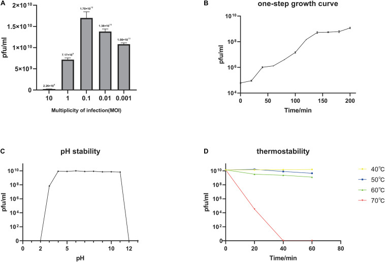
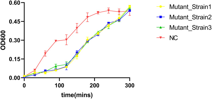
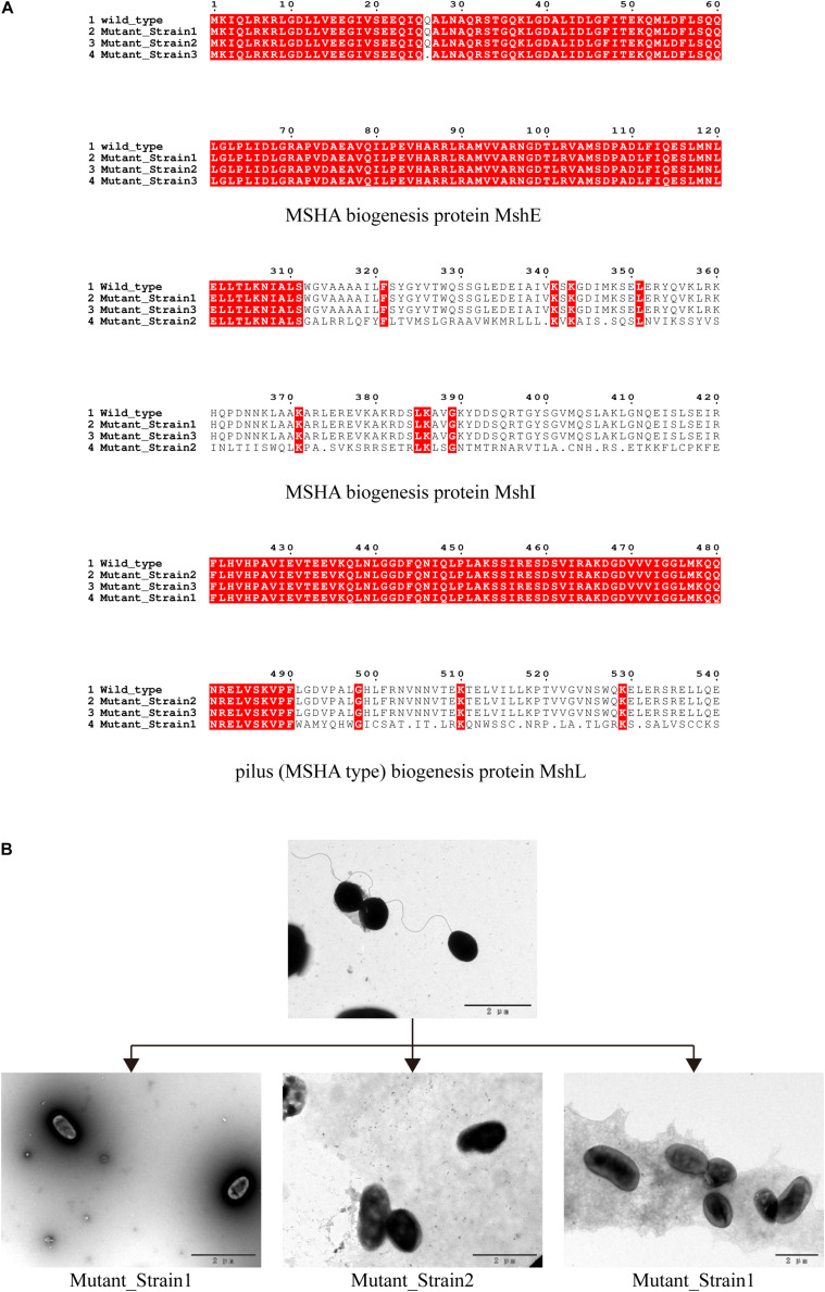
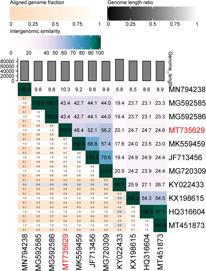
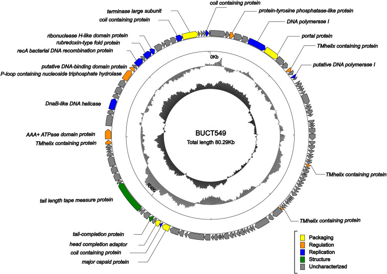
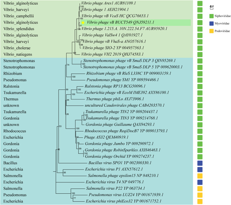
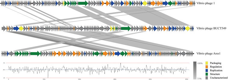
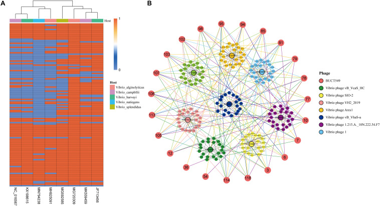
Vibrio alginolyticus is one of the most important of pathogens that can infect humans and a variety of aquatic animals, and it can cause food poisoning and septicemia in humans. Widely used antibiotics are gradually losing their usefulness, and phages are gaining more attention as new antibacterial strategies. To have more potential strategies for controlling pathogenic bacteria, we isolated a novel V. alginolyticus phage BUCT549 from seafood market sewage. It was classified as a new member of the family Siphoviridae by transmission electron microscopy and a phylogenetic tree. We propose creating a new genus for BUCT549 based on the intergenomic similarities (maximum is 56%) obtained from VIRIDIC calculations. Phage BUCT549 could be used for phage therapy due to its stability in a wide pH (3.0-11.0) range and high-temperature (up to 60°C) environment. It had a latent period of 30-40 min and a burst size of 141 PFU/infected bacterium. In the phylogenetic tree based on a terminase large subunit, BUCT549 was closely related to eight Vibrio phages with different species of host. Meanwhile, our experiments proved that BUCT549 has the ability to infect a strain of Vibrio parahaemolyticus. A coevolution experiment determined that three strains of tolerant V. alginolyticus evaded phage infestation by mutating the MSHA-related membrane protein expression genes, which caused the loss of flagellum. This research on novel phage identification and the mechanism of infestation will help phages to become an integral part of the strategy for biological control agents.

Keywords: MSHA protein; bacteriophage; genome analysis; receptor; vibrio alginolyticus.


Conflict of interest statement

FZ, HR, and QP are from Qingdao Phagepharm Bio-tech Co., Ltd. The remaining authors declare that the research was conducted in the absence of any commercial or financial relationships that could be construed as a potential conflict of interest.

Figures

FIGURE 1
FIGURE 1
Phage morphology observed using TEM.
FIGURE 2
FIGURE 2
Biological characterization of the phage BUCT549. (A) The MOI test of BUCT549. (B) The one-step growth curve of phage BUCT549, and data points show phage titers measured at 20-min intervals. (C) pH stability of BUCT549, and data points are phage titers measured after incubation of phage at different pH for 1 h. (D) Thermostability curve of BUCT549, and data points are phage titers measured after incubating the phage at different temperatures for 20, 40, and 60 min, respectively. All assays were performed in triplicate.
FIGURE 3
FIGURE 3
V. alginolyticus kill curve. V. alginolyticus was incubated with/without the addition of the phage, and the curve was plotted by measuring V. alginolyticus OD600 at 30-min intervals.
FIGURE 4
FIGURE 4
Identification of phage recognition receptors based on coevolution. (A) Mutant strains tolerant to phage BUCT549, MSHA-like proteins affected by 100% mutated site. Mutant_Strain3 mutates at the 26th amino acid of MshE protein to produce a termination codon. Mutant_Strain2 has a deletion at amino acid 312 of the MshI protein, producing a stop codon at amino acid 340. Mutant_Strain1 has a deletion at amino acid 491 of the MshL protein, producing a stop codon at amino acid position 504. (B) Electron microscopic observation of the morphology of the host V. alginolyticus (with flagella) and three tolerate strains (without flagella).
FIGURE 5
FIGURE 5
Percentage sequence similarity between phages calculated using VIDIRIC. The horizontal and vertical coordinates indicate the corresponding phage Genebank number, and the phage in this study is marked in red font.
FIGURE 6
FIGURE 6
Genome map of BUCT549. The outermost circle represents ORFs encoded in the genome with different colors representing different functions (clockwise arrow indicates the forward-reading frame, counterclockwise arrow indicates the reverse reading frame); the dark circles in the middle represent the GC content (outward indicates greater than the average GC content compared with the whole genome, and inward indicates the opposite); the innermost circle represents the GC skew (G–C/G + C. Outward indicates > 0 and inward indicates < 0).
FIGURE 7
FIGURE 7
Phylogenetic trees based on terminase large subunit, and partial phages of the Myoviridae and Podoviridae are selected as outgroups. Vibrio phages are marked in light green, and other host phages are marked in light blue. Hosts for all phages are shown on the left of the phylogenetic trees.
FIGURE 8
FIGURE 8
Multiple-sequence alignment of phage genomes. The whole genomes of Vibrio phage BUCT549, Vibrio phage 1, and Vibrio phage Ares 1, compared using Easyfig. The gray shading indicates sequence similarities between the genomes. The lower half of the graph indicates the GC content of phage BUCT549.
FIGURE 9
FIGURE 9
Analysis of genes shared by BUCT549 with other Vibrio phages. (A) The heat map shows the different Vibrio phage genes compare to BUCT549 with red representing phage sharing the gene with BUCT549 and blue representing not sharing the gene. Phage host species are colored above the heat map. (B) Relationship between the annotated genes of BUCT549 and other phage genes, the connecting lines represent the gene shared by phages.

Similar articles

Cited by

References

    1. Adriaenssens E., Brister J. R. (2017). How to Name and Classify Your Phage: an Informal Guide. Viruses 9:70. 10.3390/v9040070 - DOI - PMC - PubMed
    1. Altekruse S. F., Bishop R. D., Baldy L. M., Thompson S. G., Wilson S. A., Ray B. J., et al. (2000). Vibrio gastroenteritis in the US Gulf of Mexico region: the role of raw oysters. Epidemiol. Infect. 124 489–495. 10.1017/s0950268899003714 - DOI - PMC - PubMed
    1. Bolger A. M., Lohse M., Usadel B. (2014). Trimmomatic: a flexible trimmer for Illumina sequence data. Bioinformatics 30 2114–2120. 10.1093/bioinformatics/btu170 - DOI - PMC - PubMed
    1. Bortolaia V., Kaas R. S., Ruppe E., Roberts M. C., Schwarz S., Cattoir V., et al. (2020). ResFinder 4.0 for predictions of phenotypes from genotypes. J. Antimicrob. Chemother. 75 3491–3500. 10.1093/jac/dkaa345 - DOI - PMC - PubMed
    1. Brettin T., Davis J. J., Disz T., Edwards R. A., Gerdes S., Olsen G. J., et al. (2015). RASTtk: a modular and extensible implementation of the RAST algorithm for building custom annotation pipelines and annotating batches of genomes. Sci. Rep. 5:8365. 10.1038/srep08365 - DOI - PMC - PubMed

LinkOut - more resources