

Myeloid-Derived Suppressor Cells as a Potential Biomarker and Therapeutic Target in COVID-19
- PMID: 34220859
- PMCID: PMC8250151
- DOI: 10.3389/fimmu.2021.697405
Myeloid-Derived Suppressor Cells as a Potential Biomarker and Therapeutic Target in COVID-19
Abstract
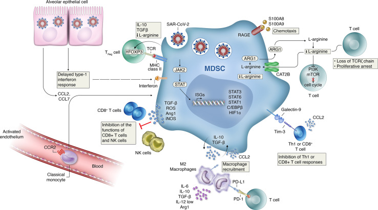
Clinical presentations of COVID-19 are highly variable, yet the precise mechanisms that govern the pathophysiology of different disease courses remain poorly defined. Across the spectrum of disease severity, COVID-19 impairs both innate and adaptive host immune responses by activating innate immune cell recruitment, while resulting in low lymphocyte counts. Recently, several reports have shown that patients with severe COVID-19 exhibit a dysregulated myeloid cell compartment, with increased myeloid-derived suppressor cells (MDSCs) correlating with disease severity. MDSCs, in turn, promote virus survival by suppressing T-cell responses and driving a highly pro-inflammatory state through the secretion of various mediators of immune activation. Here, we summarize the evidence on MDSCs and myeloid cell dysregulation in COVID-19 infection and discuss the potential of MDSCs as biomarkers and therapeutic targets in COVID-19 pneumonia and associated disease.
Keywords: COVID-19; MDSC; biomarkers; immunity; immunology.
Copyright © 2021 Rowlands, Segal and Hartl.
Conflict of interest statement
Authors MR, FS and DH were employed by the company Novartis.
Figures
Similar articles
-
Low Levels of Granulocytic Myeloid-Derived Suppressor Cells May Be a Good Marker of Survival in the Follow-Up of Patients With Severe COVID-19.Front Immunol. 2022 Jan 28;12:801410. doi: 10.3389/fimmu.2021.801410. eCollection 2021. Front Immunol. 2022. PMID: 35154077 Free PMC article.
-
Expansion of myeloid-derived suppressor cells in patients with severe coronavirus disease (COVID-19).Cell Death Differ. 2020 Nov;27(11):3196-3207. doi: 10.1038/s41418-020-0572-6. Epub 2020 Jun 8. Cell Death Differ. 2020. PMID: 32514047 Free PMC article.
-
Functional monocytic myeloid-derived suppressor cells increase in blood but not airways and predict COVID-19 severity.J Clin Invest. 2021 Mar 15;131(6):e144734. doi: 10.1172/JCI144734. J Clin Invest. 2021. PMID: 33492309 Free PMC article.
-
New Discovery of Myeloid-Derived Suppressor Cell's Tale on Viral Infection and COVID-19.Front Immunol. 2022 Feb 3;13:842535. doi: 10.3389/fimmu.2022.842535. eCollection 2022. Front Immunol. 2022. PMID: 35185933 Free PMC article. Review.
-
Myeloid-Derived Suppressor Cells in COVID-19: The Paradox of Good.Front Immunol. 2022 Apr 27;13:842949. doi: 10.3389/fimmu.2022.842949. eCollection 2022. Front Immunol. 2022. PMID: 35572540 Free PMC article. Review.
Cited by
-
Low Levels of Granulocytic Myeloid-Derived Suppressor Cells May Be a Good Marker of Survival in the Follow-Up of Patients With Severe COVID-19.Front Immunol. 2022 Jan 28;12:801410. doi: 10.3389/fimmu.2021.801410. eCollection 2021. Front Immunol. 2022. PMID: 35154077 Free PMC article.
-
Enrichment of a neutrophil-like monocyte transcriptional state in glioblastoma myeloid suppressor cells.Res Sq [Preprint]. 2023 Dec 28:rs.3.rs-3793353. doi: 10.21203/rs.3.rs-3793353/v1. Res Sq. 2023. PMID: 38234734 Free PMC article. Preprint.
-
The deciphering of the immune cells and marker signature in COVID-19 pathogenesis: An update.J Med Virol. 2022 Nov;94(11):5128-5148. doi: 10.1002/jmv.28000. Epub 2022 Jul 23. J Med Virol. 2022. PMID: 35835586 Free PMC article. Review.
-
Hallmarks of immune response in COVID-19: Exploring dysregulation and exhaustion.Semin Immunol. 2021 Jun;55:101508. doi: 10.1016/j.smim.2021.101508. Epub 2021 Oct 26. Semin Immunol. 2021. PMID: 34728121 Free PMC article. Review.
-
Reconstruction of the cell pseudo-space from single-cell RNA sequencing data with scSpace.Nat Commun. 2023 Apr 29;14(1):2484. doi: 10.1038/s41467-023-38121-4. Nat Commun. 2023. PMID: 37120608 Free PMC article.
References
-
- W. H. O . Covid-19: World Health Organization. (2021). Available at: https://www.who.int/.
Publication types
MeSH terms
Substances
LinkOut - more resources
Full Text Sources
Medical