Editorial
. 2021 Mar;16(1):117-124.
doi: 10.26574/maedica.2020.16.1.112.

Multimodality Treatment of Low-Grade Ruptured Brain Arteriovenous Malformations Presenting with Life-Threatening Intracranial Hematoma

Affiliations
Editorial

Multimodality Treatment of Low-Grade Ruptured Brain Arteriovenous Malformations Presenting with Life-Threatening Intracranial Hematoma

Vasileios Panagiotopoulos et al. Maedica (Bucur). 2021 Mar.

Abstract

Introduction:Acute management of low-grade but life-threatening ruptured arteriovenous malformations (AVM) with simultaneous hematoma evacuation remains controversial. The current report aimed to present a case series of multimodality management of low-grade (Spetzler-Martin I-II) but life-threatening ruptured arteriovenous malformations. Methods:A consecutive case series of six Spetzler-Martin (SM) grade I-II ruptured AVM patients with concurrent life-threatening hematoma initially treated with hematoma removal and, when possible, with simultaneous AVM extirpation is presented. Supplementary treatment was also applied when deemed necessary. Median clinical follow-up was 15.6 months. Neurological assessment was performed on admission (Glasgow coma scale score - GCS) and at final follow-up (modified Rankin scale score - mRS). Results:Intraparenchymal hematoma was evacuated in all six cases, with simultaneous AVM extirpation in three cases. Preoperative embolization was done in one patient, whereas postoperative embolization was performed in three additional patients. Supplementary radiosurgery was applied in one patient. Complete AVM occlusion was achieved in all patients. At the final follow-up (15.6 months), 33.3% of patients were asymptomatic, 50% had a non-significant or slight disability (mRS score 1-2), whereas one patient died. All patients with preoperative GCS score of 8 or higher had a favorable outcome. Conclusion:Acute surgical hemorrhagic clot evacuation as first step, followed by simultaneous AVM extirpation when feasible, may result in favorable clinical outcome in ruptured low-grade (SM I&II) brain AVMs with life-threatening hematoma. Embolization has a supplementary role in the acute phase of treatment either by either securing the bleeding source preoperatively or occluding the residual malformation especially in cases of technically demanding AVM removal.


Figures

TABLE 1.
TABLE 1.
Patients’ demographical and clinical data, summary of cases
FIGURE 1.
FIGURE 1.
a) Computed tomography (CT) of the brain revealing a large right frontoparietal intraparenchymal hematoma with intraventricular expansion. b) Postoperative computed tomography angiography (CTA) showing a residual arteriovenous malformation (AVM) in the right frontoparietal area. c) Digital subtraction angiography (DSA) showing a residual deep located arteriovenous malformation with arterial feeders from the right pericallosal artery. d) A single parietal Onyx injection into the residual arteriovenous malformation (AVM) up to the origin of the draining vein. e) Post-embolization (DSA) confirmed the complete AVM occlusion after a combination of surgical and endovascular techniques. f) Digital unsubtracted angiography showing the surgical clips (red arrow) and the Onyx cast (blue arrow)
FIGURE 2.
FIGURE 2.
a) Brain computed tomography (CT) scan shows a 7 cm intraparenchymal hematoma (asterisk) with significant midline shift due to a superficial arteriovenous malformation (AVM) (white arrow). b) Computed tomography angiography (CTA) showing the AVM (white arrow), a single arterial feeder (red arrow) and a single draining vein towards the superficial middle cerebral vein (blue arrow). c) Intraoperative view of the AVM (white arrow). d, e) Postoperative digital angiography of the arterial and early venous phase confirmed the complete removal of the AVM without any residual lesion. f) Postoperative CT scan demonstrates the complete excision of the arteriovenous malformation without any brain hematoma or ischemic lesion
FIGURE 3.
FIGURE 3.
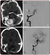
a) Brain CT scan showing a hemorrhage in the right cerebellar lobe with obstruction of the 4th ventricle and the aqueduct; b) DSA reveals a SM grade I AVM with arterial feeders from right AICA (white arrow); c, d) Postoperative CT and DSA confirmed the evacuation of the cerebellar hematoma and simultaneous complete removal of the AVM after an emergency suboccipital craniectomy
FIGURE 4.
FIGURE 4.
a) Digital subtraction angiography showing a SM grade I AVM with arterial feeders from right AICA and PICA with a low nidal aneurysm that was recognized as the source of cerebellar hemorrhage (white arrow); b-f) Superselective embolization of the ruptured nidal aneurysm was performed on an emergency basis; g) Post-embolization CT shows the cyanoacrylate cast used for embolization inside the extended cerebellar hematoma. The patient subsequently underwent surgical removal of the cerebellar hematoma and AVM, but finally died due to severe posterior fossa edema and initial brain stem injury

References

    1. Monteiro CA, Cannon G, Moubarac J-C, et al. The UN Decade of Nutrition, the NOVA food classification and the trouble with ultra-processing, Public Health Nutrition. 2018;1:5–17. - PMC - PubMed
    1. . European Lung White Book. www.erswhitebook.org, Chapter 21, p. 138.
    1. Takano H, Inone KI. Environmental pollution and allergies. J Toxicol Pathology. 2017;30:193–199. - PMC - PubMed
    1. Gupta R. JAMA Netw Open. 2019.
    1. Julia V, Macia L, Dombrowicz D. The impact of diet on asthma and allergic diseases. Nature Reviews Immunology. 2015;15:308–322. - PubMed

Publication types

LinkOut - more resources