Review
. 2021 May 18;12(24):8288-8310.
doi: 10.1039/d1sc01359a.

Recent advances in activity-based probes (ABPs) and affinity-based probes (A f BPs) for profiling of enzymes

Affiliations
Review

Recent advances in activity-based probes (ABPs) and affinity-based probes (A f BPs) for profiling of enzymes

Haixiao Fang et al. Chem Sci. .

Abstract

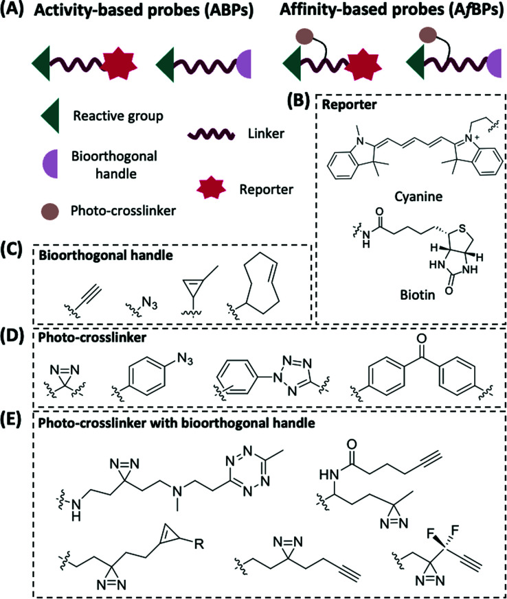
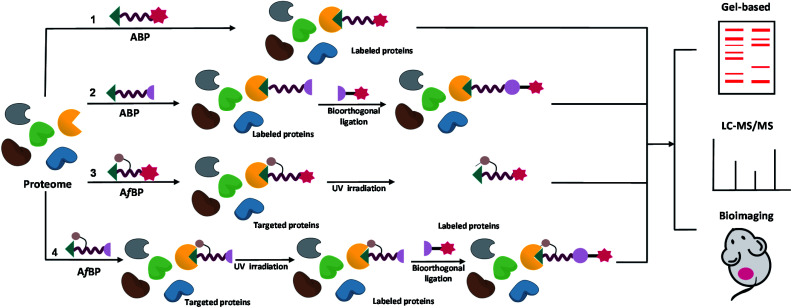
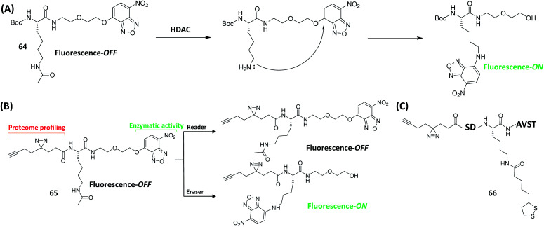
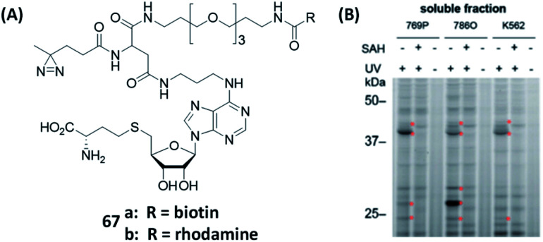
Activity-based protein profiling (ABPP) is a technique that uses highly selective active-site targeted chemical probes to label and monitor the state of proteins. ABPP integrates the strengths of both chemical and biological disciplines. By utilizing chemically synthesized or modified bioactive molecules, ABPP is able to reveal complex physiological and pathological enzyme-substrate interactions at molecular and cellular levels. It is also able to provide critical information of the catalytic activity changes of enzymes, annotate new functions of enzymes, discover new substrates of enzymes, and allow real-time monitoring of the cellular location of enzymes. Based on the mechanism of probe-enzyme interaction, two types of probes that have been used in ABPP are activity-based probes (ABPs) and affinity-based probes (AfBPs). This review highlights the recent advances in the use of ABPs and AfBPs, and summarizes their design strategies (based on inhibitors and substrates) and detection approaches.


Conflict of interest statement

There are no conflicts to declare.

Figures

Fig. 1
Fig. 1. General components of ABPs and AfBPs (A) and structures of commonly used reporter groups, bioorthogonal handles and photo-crosslinkers. (B) Examples of the reporter groups: cyanine fluorophore for visualization and biotin as an affinity tag. (C) Examples of commonly used bioorthogonal handles in ABPP. (D) Examples of photo-crosslinkers that are commonly used in ABPP. (E) Examples of photo-crosslinkers with bioorthogonal handles for AfBPs.
Fig. 2
Fig. 2. The general workflow of ABPP using ABPs and AfBPs, from identification to subsequent enrichment and quantification of target proteins. Target identification using ABPs without (1) and with (2) a bioorthogonal reporter. Target identification using AfBPs without (3) and with (4) a bioorthogonal reporter. ABP-modified proteins can be detected and identified using a variety of biochemical and cell biological techniques including gel-based fluorescence analysis, mass spectrometry-based analysis and bioimaging.
Fig. 3
Fig. 3. Kinase inhibitor-based ABPs and AfBPs and their in cellulo applications. (A) Reported kinase inhibitor-based ABPs and AfBPs. (B) Gel-based labeling of BTK with 2 in BTK-positive (Toledo, Namalwa) and BTK-negative cells (Jurkat), only positive cells showed a band around 75 kDa which demonstrated the successful labeling of cellular BTK. Reproduced from ref. with permission from Royal Society of Chemistry, copyright 2019. (C) Imaging of JNK1-GFP-transfected HeLa cells using 7a with two TP reporters, both of which successfully labeled JNK1 in living cells. Reproduced from ref. with permission from Royal Society of Chemistry, copyright 2019. (D) Reported sulfonyl fluoride-based ABPs for kinases (left). In-gel fluorescence analysis of Jurkat cells treated with either DMSO or the probe competitor, followed by treatment with 11a to 11c. 11 was successfully used for broad-spectrum kinase labeling in cells. P = probe, C = competitor. Reproduced from ref. with permission from American Chemical Society, copyright 2017.
Fig. 4
Fig. 4. Two qABP working strategies. (A) Schematic illustration of activity-dependent fluorescent labeling of Cys proteases by using qABP. (B) Another strategy of the enzyme activity-dependent qABP reaction. The structure of qABP contains a mandelic acid core which was surrounded by a fluorophore (red), a quencher and an enzyme WH (blue). The resulting intermediate of enzymatic reaction has three possible pathways within the cells: (pathway 1) hydration/quenching by water; (pathway 2) covalent attachment to the target enzyme; (pathway 3) accumulation in the organelle where the enzymatic reaction occurs. Reproduced from ref. with permission from American Chemical Society, copyright 2011.
Fig. 5
Fig. 5. The working strategies and structures of the “AND-gate” probes. (A) Schematic illustration of an AND-gate fluorescent probe. Q = quencher, F = fluorophore. Reproduced from ref. with permission from Springer Nature, copyright 2020. (B) Structures of reported AND-gate probes.
Fig. 6
Fig. 6. Protease qABPs for visual labeling of different proteases in live cells and mouse tumors. (A) Structures of reported ABPs to study proteases. (B) In-gel fluorescence analysis of Casp3 in MM1s cells treated with 14. The addition of Casp3 inhibitor blocked the labeling of Casp3 by the probe, which demonstrated the specificity of the probe to Casp3. Reproduced from ref. with permission from Royal Society of Chemistry, copyright 2016. (C) Labeling of cathepsin B in non-small lung cancer cells from patient using 15 and anti-cathepsin B FITC antibody. The labeling of cathepsin B using 15 was blocked by the addition of cathepsin B inhibitor, but the inhibitor did not influence the labeling efficiency of the antibody. Reproduced from ref. with permission from Royal Society of Chemistry, copyright 2019. (D) Still images from Novadaq Spy Elite and SurgVision Explorer Air of 4T1 tumors at different time points after injection of 16, which showed the potential of 16. Reproduced from ref. with permission from Springer, copyright 2020. (E) Specific labeling of active MMP-14 using protein engineering coupled with 17. The ABP only labeled active MMPs and cannot label enzymes that are inaccessible to zinc or their inhibitory forms. Reproduced from ref. with permission from American Chemical Society, copyright 2018.
Fig. 7
Fig. 7. Inhibitor-based ABPs for glycosidases and labeling of GBA and hrGBA in human tissues by using ABPs. (A) Structures of cyclophellitol-derived epoxides- and aziridines-based ABPs. (B) Structures of various reporter groups used in ABPs. (C) Overlay of electron micrographs and confocal fluorescence images of NHDF ultrathin cryosections expressing Man-R. The metabolic processes of hrGBA and endogenous GBA in vivo were traced using 18. 18a-labeled hrGBA (green), 18b-labeled endogenous GBA (red) and nuclei labeled with DAPI (blue). E = endosome; L = lysosome. M = mitochondria; PM = plasma membrane. Reproduced from ref. with permission from Wiley-VCH GmbH, copyright 2019.
Fig. 8
Fig. 8. Inhibitor-based ABPs for labeling of six human proteasome subunits and their working strategies. (A) Structures of ABPs that target different subunits of human proteasomes. (B) Schematic representation of ABPP using proteasome ABP mixture. Visual gel analysis of six human proteasome subunits was achieved using three ABP mixtures. Reproduced from ref. with permission from Wiley-VCH GmbH, copyright 2016.
Fig. 9
Fig. 9. ABPs and AfBPs designed based on specific enzyme inhibitors and their labeling of enzymes in cells. (A) Structures of reported enzyme inhibitor-based ABPs. (B) Confocal images of S. aureus Newman labeled with 35 (1). Confocal images of S. aureus Newman-GFP cell labeled with 35 during exponential phase (2 and 3) and stationary phase (4 and 5). 35 successfully demonstrated that FphB is concentrated in the isolated membrane of bacterial cells. Reproduced from ref. with permission from Springer Nature, copyright 2018. (C) Proteome profiles of MCF-7 cells labeled with 39, with a visible FL band around 55 kDa. These demonstrated the specific labeling of PHGDH by 39 in living cells. (D) Confocal images of MCF-7 cells treated with 39 and anti-PHGDH. Reproduced from ref. with permission from Wiley-VCH GmbH, copyright 2018.
Fig. 10
Fig. 10. A bisubstrate probe strategy for specific labeling of UGTs. (A) The structures of steviol and the reported substrate-based ABP and AfBPs. (B) Bisubstrate probe strategy for the identification of UGT cellular localization and activities by steviol-based ABPs. Reproduced from ref. with permission from Royal Society of Chemistry, copyright 2020.
Fig. 11
Fig. 11. The mechanism and application of NAD+-based ABPs (modified adenine moieties as an example). Upon the polyADP-ribosylation via PARPs, the PAR is formed from a single ADP-ribosyl linkage on the substrate proteins, which is usually followed by the enrichment and gel-/mass spectrometry-based analysis of PARP substrate proteins through click chemistry.
Fig. 12
Fig. 12. Structures of reported ABPs based on NAD+ by modifying adenine or nicotinamide ribose-OH at different sites.
Fig. 13
Fig. 13. Gel-based analysis of substrate activities of NAD+, 48b, f and 45b, c for (A) PARP1; (B) PARP2; (C) catalytic domain of PARP5a; and (D) catalytic domain of PARP10. All the probes were successfully recognized by the corresponding enzymes, and the addition of PARP inhibitor olaparib efficiently inhibited the interactions between the probes and PARPs. Reproduced from ref. with permission from Springer Nature, copyright 2019.
Fig. 14
Fig. 14. Two approaches of the “bump-hole” strategy for identifying PARP target proteins. (A) Targeting a conserved residue in the nicotinamide binding site of the PARP catalytic domain. (B) Targeting a conserved residue in the adenine binding site of the PARP catalytic domain.
Fig. 15
Fig. 15. Use clickable AfBPs to capture proteins associated with PARylation. (A) Structures of reported bifunctional NAD+ probes. (B) Schematic illustration of the process for capturing PARylation-related interacting proteins using a bifunctional NAD+ probe. Reproduced from ref. with permission from American Chemical Society, copyright 2021. (C) Structures of reported BAD-based clickable AfBPs.
Fig. 16
Fig. 16. Ub-based ABPs and AfBPs for DUBs and E1, E2, E3 enzyme studies. (A) Structures of reported ABPs and AfBPs based on Ub. (B) Mechanism of 62 for the labeling of E1, E2 and E3 enzymes. Pathway (1): enzymatic covalent binding process, pathway (2): natural transtrans thioesterification process.
Fig. 17
Fig. 17. Single-step fluorescence detection and identification of enzyme readers and erasers using AfBPs. (A) Schematic of HDAC detection using fluorometric method. (B) Schematic illustration of 65 for detecting enzymatic activity and proteomic profiling studies. Single-step fluorescence detection and proteomic profiling of HDAC can be achieved simultaneously by using 65. (C) Structure of AfBP 66 for the identification of Klip erasers.
Fig. 18
Fig. 18. Gel-based analysis of MTs was performed using AfBPs. (A) Structures of reported AfBPs for MTs. (B) Gel-based profiling with 67b in human renal cancer cell (769P and 786O) and human leukemia cell (K562) lysates. 67 successfully labeled MTs in a variety of cancer cells. The addition of SAH (natural substrate) or the lack of UV irradiation resulted in the loss of MT labeling. Reproduced from ref. with permission from American Chemical Society, copyright 2019.

Similar articles

Cited by

References

    1. Markel U. Essani K. D. Besirlioglu V. Schiffels J. Streit W. R. Schwaneberg U. Chem. Soc. Rev. 2020;49:233–262. - PubMed
    1. Wijdeven R. H. Neefjes J. Ovaa H. Trends Cell Biol. 2014;24:751–760. - PubMed
    1. Uttamchandani M. Lu C. H. Yao S. Q. Acc. Chem. Res. 2009;42:1183–1192. - PubMed
    1. Evans M. J. Cravatt B. F. Chem. Rev. 2006;106:3279–3301. - PubMed
    1. Sanman L. E. Bogyo M. Annu. Rev. Biochem. 2014;83:249–273. - PubMed