

Delayed presentation of a traumatic scalp arteriovenous fistula
- PMID: 34221569
- PMCID: PMC8247668
- DOI: 10.25259/SNI_263_2021
Delayed presentation of a traumatic scalp arteriovenous fistula
Abstract
Background: Arteriovenous (AV) fistulas of the scalp are extracranial vascular malformations commonly caused by trauma and typically present within 3 years. Although they follow a benign course, they can be esthetically displeasing.
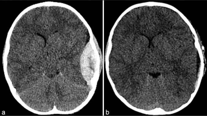
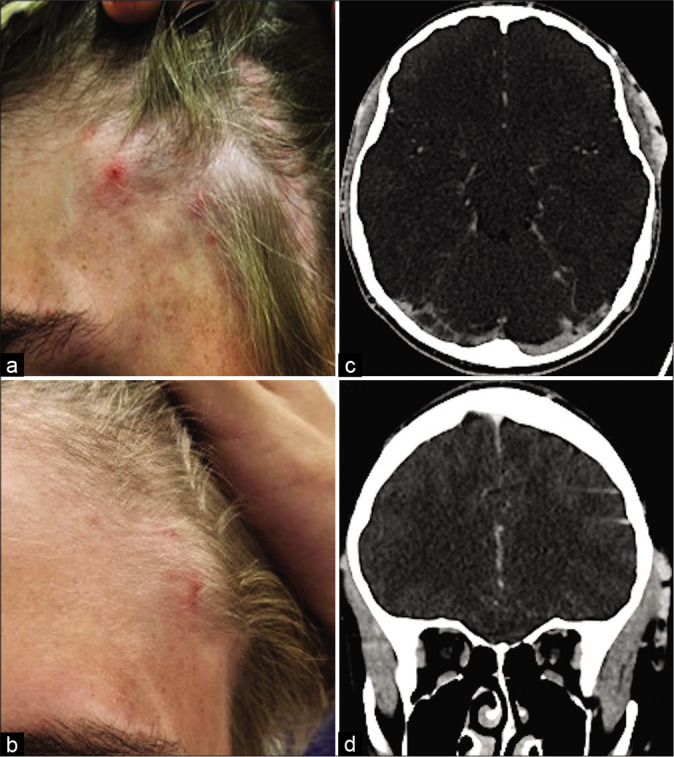
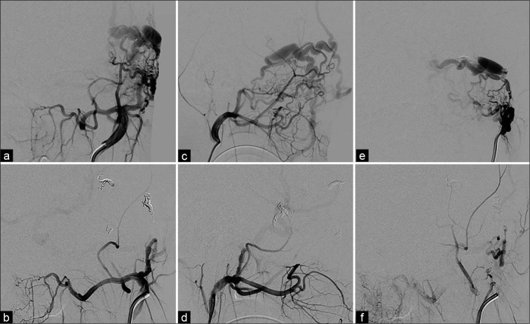
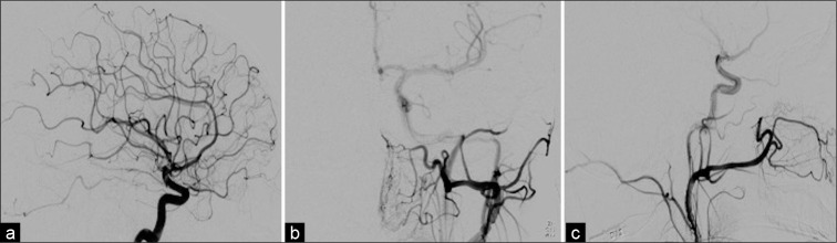
Case description: We present an atypical onset of scalp AV fistula in a patient with a 1-year history of the left-sided pulsatile tinnitus and scalp swelling 7 years after a traumatic epidural hematoma evacuation. Our patient was found to have an 8 mm AV fistula supplied by the deep temporal artery. Endovascular embolization was performed using eight coils. There was no complication from the procedure, and the patient's pulsatile tinnitus and swelling resolved immediately after embolization. Follow-up angiogram demonstrated complete obliteration of the AV fistula.
Conclusion: Delayed presentation of traumatic scalp AV fistula is very rare, and it is important to keep this in the differential in patients with scalp swelling after head trauma.
Keywords: Arteriovenous fistula; Head trauma; Iatrogenic fistula; Neuroendovascular.
Copyright: © 2021 Surgical Neurology International.
Conflict of interest statement
There are no conflicts of interest.
Figures
References
-
- Fisher-Jeffes ND, Domingo Z, Madden M, de Villiers JC. Arteriovenous malformations of the scalp. Neurosurgery. 1995;36:656–60. discussion 660. - PubMed
-
- Gurkanlar D, Gonul M, Solmaz I, Gonul E. Cirsoid aneurysms of the scalp. Neurosurg Rev. 2006;29:208–12. - PubMed
-
- Leke B, Peter R. Traumatic arteriovenous fistula of the scalp. J Neurosurg. 1987;66:773–4. - PubMed
-
- Luessenhop A. Cirsoid aneurysms of the scalp. J Neurosurg. 1991;75:167. - PubMed
Publication types
LinkOut - more resources
Full Text Sources