. 2021 May 31:12:249.
doi: 10.25259/SNI_532_2020. eCollection 2021.

Magnetic resonance imaging analysis of human skull diploic venous anatomy

Affiliations

Magnetic resonance imaging analysis of human skull diploic venous anatomy

Abdullah Alarfaj et al. Surg Neurol Int. .

Abstract

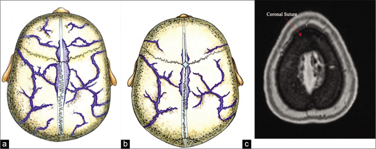
Background: The skull diploic venous space (DVS) represents a potential route for cerebrospinal fluid (CSF) diversion and absorption in the treatment of hydrocephalus. The goal of this study was to carry out a detailed characterization of the drainage pattern of the DVS of the skull using high-resolution MRI, especially the diploic veins draining to the lacunae laterales (LLs) since the LLs constitute an important channel for the CSF to access the superior sagittal sinus and subsequently the systemic circulation. The objective was to identify those skull regions optimally suited for an intraosseous CSF diversion system.

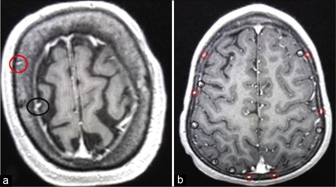
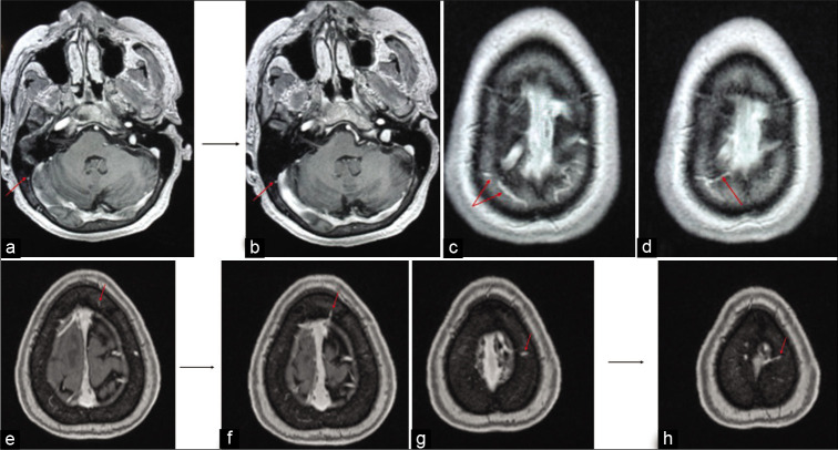
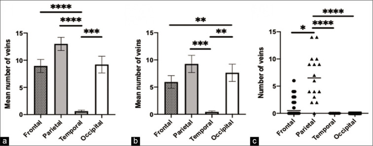
Methods: High-resolution, T1-weighted MRI scans from 20 adult and 16 pediatric subjects were selected for analysis. Skulls were divided into four regions, that is, frontal, parietal, temporal, and occipital. On each scan, a trained observer counted all diploic veins in every skull region. Each diploic vein was also followed to determine its final drainage pathway (i.e., dural venous sinus, dural vein, LL, or indeterminate).

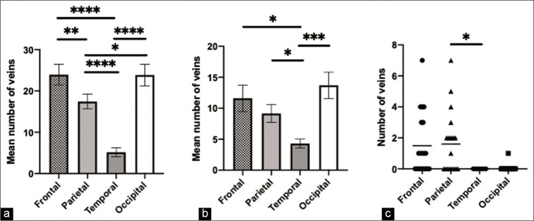
Results: In the adult age group, the frontal and occipital skull regions showed the highest number of diploic veins. However, the highest number of draining diploic veins connecting to the lacunae lateralis was found in the frontal and parietal skull region, just anterior and just posterior to the coronal suture. In the pediatric age group, the parietal skull region, just posterior to the coronal suture, showed the highest overall number of diploic veins and also the highest number of draining diploic veins connecting to the LL.

Conclusion: This study suggested that diploic venous density across the skull varies with age, with more parietal diploic veins in the pediatric age range, and more occipital and frontal diploic veins in adults. If the DVS is ultimately used for CSF diversion, our anatomical data point to optimal sites for the insertion of specially designed intraosseous infusion devices for the treatment of hydrocephalus. Likely the optimal sites for CSF diversion would be the parietal region just posterior to the coronal suture in children, and in adults, frontal and/or parietal just anterior or just posterior to the coronal suture.

Keywords: Cerebrospinal fluid diversion; Diploic venous space; Skull regions.


Conflict of interest statement

There are no conflicts of interest.

Figures

Figure 1:
Figure 1:
The definition of skull regions used in the data analysis based on the skull sutures. The frontal skull region was defined as the region of the skull anterior to the coronal suture and superior to the sphenofrontal sutures. The parietal skull region was defined as the region of the skull posterior to the coronal suture and superior to the squamosal suture and anterior/superior to the lambdoid sutures. The temporal skull region was defined as region of the skull inferior to the squamosal and sphenofrontal sutures and anterior to the occipitomastoid suture. The occipital skull region was defined as the region of the skull inferior to the lambdoid suture and posterior to the occipitomastoid suture. Source https://openstax.org/books/anatomy-andphysiology/pages/7-2-the-skull. licensed under the Creative Commons Attribution 3.0 Unported license; https://creativecommons.org/licenses/by/3.0/deed.en. Additional naming designation of the sutures was made.
Figure 2:
Figure 2:
Illustration of the diploic veins versus cortical veins. The red circles in A and B are identifying the veins coursing in the diploic venous space. Each diploic vein was counted in each skull region and was followed to identify the ultimate pathway. Cortical veins were not counted but are encircled with black circles for illustration.
Figure 3:
Figure 3:
Illustrations of the different pathways for the diploic veins. The MRI in (a and b) shows a diploic vein in the right occipital region marked with red arrow appearing to drain directly into the transvers sinus. The MRI in (c and d) shows diploic veins marked with red arrow in the right parietal region which appear to drain into the lateral aspect of the superior sagittal sinus. The MRI in (e and f) shows a diploic vein in the left frontal region marked with red arrow appearing to drain into the superior sagittal sinus. The MRI in (g and h) shows a diploic vein in the left parietal region marked with red arrow appearing to drain into the lacunae laterales.
Figure 4:
Figure 4:
Adult age group findings. (a) The comparison and significant difference in the mean number of veins in the four skull regions, (b) the comparison and significant difference in the mean number of draining veins in the four skull regions, (c) the comparison and significant difference in the number of veins draining to the lacunae laterales in the four skull regions. * = p<0.05, ** = p<0.01, *** = p<0.001, **** = p<0.0001.
Figure 5:
Figure 5:
Pediatric age group findings. (a) The comparison and significant difference in the mean number of veins in the four skull regions, (b) the comparison and significant difference in the mean number of draining veins in the four skull regions, (c) the comparison and significant difference in the number of veins draining to the lacunae laterales in the four skull regions. * = p<0.05, ** = p<0.01, *** = p<0.001, **** = p<0.0001.
Figure 6:
Figure 6:
The Lacuane Laterales in relation to the coronal suture and skull diploic veins. The illustrative pictures in A and B shows the anatomic location of the LL just adjacent to the coronal suture which corresponds with the radiological finding of the highest number of diploic veins draining to the LL just anterior and just posterior to the coronal suture, in the adult age group and the pediatric age group, respectively. Please refer to Rhoton Cranial Anatomy and Surgical Approaches text book figure 4.14 a page 179 and figure 4.3 c page 164 for cadaveric dissection showing LL in relation to coronal suture. (a) Illustration showing the LL in relation to the coronal suture in adults. (b) Illustration showing the LL in relation to the coronal suture in pediatrics. (c) MRI MP RAGE showing the LL in relation to the coronal suture “red arrow pointing at the coronal suture.”

References

    1. Aschoff A, Kremer P, Hashemi B, Kunze S. The scientific history of hydrocephalus and its treatment. Neurosurg Rev. 1999;22:67–93. discussion 94-5. - PubMed
    1. Baert EJ, Dewaele F, Vandersteene J, Hallaert G, Kalala JO, van Roost D. Treating hydrocephalus with retrograde ventriculosinus shunt: Prospective clinical study. World Neurosurg. 2018;118:e34–42. - PubMed
    1. Brinker T, Stopa E, Morrison J, Klinge P. A new look at cerebrospinal fluid circulation. Fluids Barriers CNS. 2014;11:10. - PMC - PubMed
    1. Bulleid LS, Hughes T, Bhatti I, Leach PA. Low-pressure headaches following foramen magnum decompression secondary to absorption of cerebrospinal fluid into the venous system of the diploic space. Childs Nerv Syst. 2016;32:897–9. - PubMed
    1. Fox RJ, Walji AH, Mielke B, Petruk KC, Aronyk KE. Anatomic details of intradural channels in the parasagittal dura: A possible pathway for flow of cerebrospinal fluid. Neurosurgery. 1996;39:84–90. discussion 90-1. - PubMed

LinkOut - more resources