Sclerosing Encapsulating Peritonitis: A Rare Cause of Intestinal Obstruction
- PMID: 34221753
- PMCID: PMC8237918
- DOI: 10.7759/cureus.15291
Sclerosing Encapsulating Peritonitis: A Rare Cause of Intestinal Obstruction
Abstract
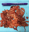
Sclerosing encapsulating peritonitis (SEP) is a rare clinical entity that may cause small bowel obstruction. It is characterized by a thick fibrocollagenous cocoon-like membrane. Surgical intervention is the mainstay of treatment. A 36-year-old Pakistani man presented with recurrent attacks of colicky abdominal pain, distention, vomiting, and constipation. Abdominal CT revealed a thick enhanced membrane forming a sac that contained clusters of small intestinal loops. Exploratory laparotomy showed a thick membrane containing the small bowel and extensive inter-loop adhesions. The sac underwent decortication and excision, inter-loop adhesions were released, and an appendectomy was performed. The patient tolerated the procedure and was discharged in good condition.
Keywords: abdominal cocoon; abdominal pain; bowel obstruction; case report; sclerosing encapsulating peritonitis.
Copyright © 2021, Yusuf et al.
Conflict of interest statement
The authors have declared that no competing interests exist.
Figures


References
-
- Peritonitis chronic fibrosa incapsulata. Owtschinnikow PJ. Arch Klin Chir. 1907;83:623–634.
-
- Unusual small intestinal obstruction in adolescent girls: the abdominal cocoon. Foo KT, Ng KC, Rauff A, Foong WC, Sinniah R. Br J Surg. 1978;65:427–430. - PubMed
-
- Abdominal cocoon: multi-detector row CT with multiplanar reformation and review of literatures. Wang Q, Wang D. Abdom Imaging. 2010;35:92–94. - PubMed
-
- Abdominal cocoon in children: a report of four cases. Sahoo SP, Gangopadhyay AN, Gupta DK, Gopal SC, Sharma SP, Dash RN. https://pubmed.ncbi.nlm.nih.gov/8811577. J Pediatr Surg. 1996;31:987–988. - PubMed
Publication types
LinkOut - more resources
Full Text Sources
