Review
. 2021 Jun;11(6):1365-1378.
doi: 10.1016/j.apsb.2021.03.027. Epub 2021 Apr 24.

Role of CD8+ T lymphocyte cells: Interplay with stromal cells in tumor microenvironment

Affiliations
Review

Role of CD8+ T lymphocyte cells: Interplay with stromal cells in tumor microenvironment

Qin Xie et al. Acta Pharm Sin B. 2021 Jun.

Abstract

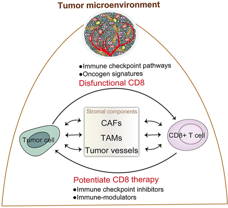
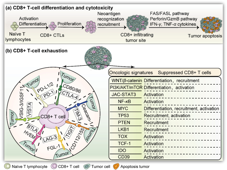
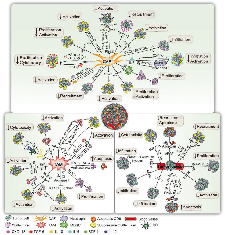
CD8+ T lymphocytes are pivotal cells in the host response to antitumor immunity. Tumor-driven microenvironments provide the conditions necessary for regulating infiltrating CD8+ T cells in favor of tumor survival, including weakening CD8+ T cell activation, driving tumor cells to impair immune attack, and recruiting other cells to reprogram the immune milieu. Also in tumor microenvironment, stromal cells exert immunosuppressive skills to avoid CD8+ T cell cytotoxicity. In this review, we explore the universal function and fate decision of infiltrated CD8+ T cells and highlight their antitumor response within various stromal architectures in the process of confronting neoantigen-specific tumor cells. Thus, this review provides a foundation for the development of antitumor therapy based on CD8+ T lymphocyte manipulation.

Keywords: Antitumor; CD8+ T lymphocyte; Immunosuppression; Immunotherapy; Stromal cell; Tumor microenvironment.


Figures

Image 1
Graphical abstract
Figure 1
Figure 1
Infiltration of CD8+ T cells into tumors: differentiation, cytotoxicity, and dysfunction. (a) CD8+ T cells are programmed into CTLs after their activation and proliferation within the TME. The tumor cells are eventually eliminated, depending on IFN-γ and TNF-α secretion and apoptosis pathways. (b) Left: Various immune checkpoints in the divided region involved in CD8+ T cell dysfunction. Once the related ligand–receptor bindings are activated, CD8+ T cells are disabled and lose killing efficacy. Right: Oncologic signatures for suppressing tumor-infiltrating CD8+ T cells. Alternatively, WNT/β-catenin inhibits the differentiation and recruitment of CD8+ T cells against tumors. Other pathways include PI3K, STAT3, NF-κB, MYC, TP53, PTEN, LKB1, TOX, TCF-1, IDO, and CD39.
Figure 2
Figure 2
Suppressive immunization regulation of CD8+ T cells with stromal cells in the TME. The suppressive immunization regulation of CD8+ T cells for pro-tumoral microenvironment with TME-related stromal components is depicted in three parts: CAFs, TAMs, and tumor vessels. (1) For CAFs, the immune checkpoint molecules CTLA-4, TIM-3, PD-1, LAG-3, and CD73 are induced to attenuate CD8+ T cells. NF-κB prevents CD8+ T cells by upregulating CXCL12. IL-6 is secreted to downregulate CD8+ T cell infiltration and IL-6/STAT3 can master the PD-1/PD-L1 pathway to impair T cells by upregulating CXCR7. Tumor-specific CD8+ T cells are inhibited by TGF-β, along with two auto-stimulatory signaling of TGF-β and SDF-1. The CXCL12/CXCR4 axis induces FAP to diminish CD8+ T cells. FAS/FASL on T cells leads to CD8+ T cell apoptosis. CD8+ T cells are excluded with HDAC6 to activate STAT3 by targeting COX2. CAF-driven ROS, Chi3L1, βig-h3, and arginase II are capable of impairing CD8+ T cell activity. (2) For the TME, the immune checkpoint pathways PD-1/PD-L1/2 and CTLA-4/CD80/86 are observed on TAMs to confine CD8+ T cell initiation. NF-κB P65 is validated to impair CD8+ CTLs by inducing B7–H4/B7S1 and anti-apoptosis gene, as well as activating PD-1. Arginase and NO activity are modulators responsible for CD8+ T cell apoptosis via IFN-γ and TNF-α. IL-10 limits cytotoxic CD8+ T cells by suppressing DC-driven IL-12 or selectively reducing B7 upregulation. In addition, STAT1, CSF1, HLA-G, HLA-E, arginase I, and SHH from macrophages are crucial regulators to deplete the CD8+ T cell response. (3) During neovascularization, VEGF inhibits CD8+ T cell homing and induces apoptosis. HIF-1α may modulate vascularization via VEGF-A. FASL is selectively expressed in tumor-driven vasculatures to hinder CD8+ T cells with soluble VEGF-A, IL-10, and PGE2. NF-κB is capable of activating FASL and downregulating cFLIP for apoptosis. Intratumoral CD8+ T cells are rejected by RGS5, resulting in the formation of abnormal blood vessels and hypoxia. HIF-1α, ETBR, B7–H3 β-AR, PDPN, and TNF-α are also pivotal vascular molecules, impeding CD8+ T cell penetration into tumor sites.

References

    1. Quail D.F., Joyce J.A. Microenvironmental regulation of tumor progression and metastasis. Nat Med. 2013;19:1423–1437. - PMC - PubMed
    1. Wellenstein M.D., Visser K.E. Cancer-cell-intrinsic mechanisms shaping the tumor immune landscape. Immunity. 2018;48:399–416. - PubMed
    1. Kim J.M., Chen D.S. Immune escape to PD-L1/PD-1 blockade: seven steps to success (or failure) Ann Oncol. 2016;27:1492–1504. - PubMed
    1. Liu Y., Zeng G. Cancer and innate immune system interactions: translational potentials for cancer immunotherapy. J Immunother. 2012;35:299–308. - PMC - PubMed
    1. Ascierto P.A., Lewis K.D., Giacomo A.M., Demidov L., Mandala M., Bondarenko I. Prognostic impact of baseline tumour immune infiltrate on disease-free survival in patients with completely resected, BRAF(v600) mutation-positive melanoma receiving adjuvant vemurafenib. Ann Oncol. 2020;31:153–159. - PubMed