Review
. 2021 Jun;11(6):1493-1512.
doi: 10.1016/j.apsb.2020.12.014. Epub 2020 Dec 19.

Extracellular vesicle activities regulating macrophage- and tissue-mediated injury and repair responses

Affiliations
Review

Extracellular vesicle activities regulating macrophage- and tissue-mediated injury and repair responses

Qian Hu et al. Acta Pharm Sin B. 2021 Jun.

Abstract

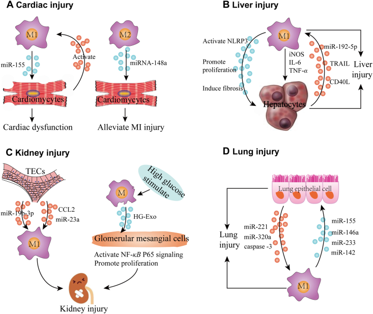
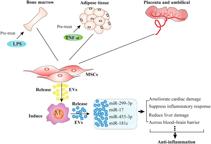
Macrophages are typically identified as classically activated (M1) macrophages and alternatively activated (M2) macrophages, which respectively exhibit pro- and anti-inflammatory phenotypes, and the balance between these two subtypes plays a critical role in the regulation of tissue inflammation, injury, and repair processes. Recent studies indicate that tissue cells and macrophages interact via the release of small extracellular vesicles (EVs) in processes where EVs released by stressed tissue cells can promote the activation and polarization of adjacent macrophages which can in turn release EVs and factors that can promote cell stress and tissue inflammation and injury, and vice versa. This review discusses the roles of such EVs in regulating such interactions to influence tissue inflammation and injury in a number of acute and chronic inflammatory disease conditions, and the potential applications, advantage and concerns for using EV-based therapeutic approaches to treat such conditions, including their potential role of drug carriers for the treatment of infectious diseases.

Keywords: ADSCs, adipose-derived stem cells; AKI, acute kidney injury; ALI, acute lung injury; AMs, alveolar macrophages; BMSCs, bone marrow stromal cells; CLP, cecal ligation and puncture; DSS, dextran sodium sulphate; EVs, extracellular vesicles; Extracellular vesicles; HSPA12B, heat shock protein A12B; HUCMSCs, human umbilical cord mesenchymal stem cells; IBD, inflammatory bowel disease; ICAM-1, intercellular adhesion molecule 1; IL-1β, interleukin-1β; Inflammatory disease; Interaction loop; KCs, Kupffer cells; KLF4, krüppel-like factor 4; LPS, lipopolysaccharides; MHC, major histocompatibility complex; MSCs, mesenchymal stromal cells; MVs, microvesicles; Macrophage; PEG, polyethylene glycol; PMFA, 5,7,30,40,50-pentamethoxyflavanone; PPARγ, peroxisome proliferator-activated receptor γ; SIRPα, signal regulatory protein α; Sepsis; Stem cell; TECs, tubular epithelial cells; TNF, tumor necrosis factor; TRAIL, tumor necrosis factor-related apoptosis-inducing ligand; Targeted therapy; Tissue injury; iNOS, inducible nitrogen oxide synthase.


Conflict of interest statement

The authors have no conflicts of interest to declare.

Figures

Image 1
Graphical abstract
Figure. 1
Figure 1
Macrophage polarization and phenotypes. Stimulation by TLR receptor signaling or by exposure to IL-4, IL-10, IL-13 and TGF-β induces macrophages to adopt M1 or M2 phenotypes, respectively, which causes these macrophage subtypes to secrete distinct sets of cytokines and chemokines that exert pro-inflammatory or anti-inflammatory effects.
Figure 2
Figure 2
EV-mediated interactions between tissue macrophages and adjacent cells can induce tissue inflammation and injury. (A) Cardiac injury. M1 macrophages secrete extracellular vesicles (EVs) containing miR-155 that induce cardiomyocyte dysfunction, while EVs secreted by injured cardiomyocytes can activate macrophages. Conversely, exosomes secreted by M2 macrophages carry miRNA-148a, which can target cardiomyocytes and reduce myocardial ischemia/reperfusion injury. (B) Liver injury. EVs secreted by M1 macrophages can activate the hepatocyte NLRP3 inflammasome pathway and promote hepatocyte proliferation, liver fibrosis, and the secretion of inflammatory factors, including iNOS, IL-6 and TNF-α, to cause liver injury. Stressed hepatocytes can secrete EV containing miR-192-5p, TRAIL and CD40L to promote M1 macrophage polarization. (C) Kidney injury. Tubular epithelial cells (TECs) secrete EVs enriched with miR-19b-3p, CCL2 and miR-23a that promote M1 macrophage polarization to induce tubulointerstitial inflammation. Conversely, M1 macrophages stimulated by high glucose secrete EVs that can activate NF-κB P65 signaling and promote proliferation of glomerular mesangial cells to cause kidney injury. (D) Lung injury. EVs secreted by lung epithelial cells carry miR-221, miR320a and caspase-3 to promote alveolar macrophages to adopt an M1 phenotype, while M1 macrophage EVs containing miR-155, miR-146a, miR-233 and miR-142 induce lung epithelial cell injury.
Figure 3
Figure 3
M1 macrophage EV interactions in inflammatory diseases affecting different tissues. EVs in the peripheral circulation carry miR-155 that activates p38/ERK signaling pathways and M1 macrophage polarization. M1 macrophage EVs (M1-EVs) demonstrate multiple pro-inflammatory effects, as they: induce proinflammatory IL-1β and TNF-α secretion in colon tissue, and endothelial dysfunction, to promote inflammatory bowel diseases (IBDs); activate NF-κB and NLRP3 signaling in acinar cells to induce acute pancreatitis (AP); target lung and kidney tissues to induce acute lung injury (ALI) and kidney injury (AKI); carry miR-34a, which upregulates TGF-β and WNT/β-catenin signaling in adipocytes to promote in insulin resistance (IR). Colon cells and adipocytes experiencing inflammatory conditions also release EVs that can promote macrophage polarization to pro-inflammatory phenotypes.
Figure 4
Figure 4
Extracellular vesicles (EVs) secreted by mesenchymal stromal cells (MSCs) from bone marrow, and adipose and umbilical cord tissue induce M2 macrophage polarization via specific miRNA cargoes to regulate an anti-inflammatory effects.

References

    1. van Furth R., Cohn Z.A., Hirsch J.G., Humphrey J.H., Spector W.G., Langevoort H.L. The mononuclear phagocyte system: a new classification of macrophages, monocytes, and their precursor cells. Bull World Health Organ. 1972;46:845–852. - PMC - PubMed
    1. Ginhoux F., Greter M., Leboeuf M., Nandi S., See P., Gokhan S. Fate mapping analysis reveals that adult microglia derive from primitive macrophages. Science. 2010;330:841–845. - PMC - PubMed
    1. Schulz C., Gomez Perdiguero E., Chorro L., Szabo-Rogers H., Cagnard N., Kierdorf K. A lineage of myeloid cells independent of Myb and hematopoietic stem cells. Science. 2012;336:86–90. - PubMed
    1. Ovchinnikov D.A. Macrophages in the embryo and beyond: much more than just giant phagocytes. Genesis. 2008;46:447–462. - PubMed
    1. Epelman S., Lavine K.J., Randolph G.J. Origin and functions of tissue macrophages. Immunity. 2014;41:21–35. - PMC - PubMed