Two-Dimensional Transesophageal Echocardiography Assessment of the Major Aortic Annulus Diameter in Patients Undergoing Transcatheter Aortic Valve Replacement
- PMID: 34221882
- PMCID: PMC8230165
- DOI: 10.4103/jcecho.jcecho_110_20
Two-Dimensional Transesophageal Echocardiography Assessment of the Major Aortic Annulus Diameter in Patients Undergoing Transcatheter Aortic Valve Replacement
Abstract
Background: Multidetector computed tomography (MDCT) is the gold standard in annulus sizing before transcatheter aortic valve replacement (TAVR). However, MDCT has limited applicability in specific subgroups of patients, such as those with atrial fibrillation and chronic kidney disease. Two-dimensional transesophageal echocardiography (2DTEE) has traditionally been limited to the long-axis measurement of the anteroposterior diameter of the aortic annulus. We describe a new 2DTEE approach for the measurement of the major diameter of the aortic annulus.
Methods: Seventy-six patients with symptomatic severe aortic valve stenosis and high surgical risk underwent MDCT and 2DTEE before TAVR. A modified five-chamber view was used to measure the major aortic annulus diameter. This was obtained starting from a mid-esophageal four chamber and retracting the TEE probe up until the left ventricular outflow tract and the left and noncoronary aortic cusps were visualized: major aortic annulus diameter was measured as the distance between their insertion points in systole.
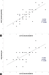
Results: Major aortic annulus diameters measured at 2DTEE showed good correlation with MDCT diameter (r = 0.79; P < 0.001) and perimeter (r = 0.87; P < 0.0001). Using factsheet-derived sizing criteria, 2DTEE alone would have allowed accurate sizing in 75% of patients, with 21% of oversizing predominantly with smaller annuli.
Conclusions: We describe a new method for 2DTEE measurement of the major aortic annulus diameter; this approach is simple, correlates with MDCT, and allows adequate TAVR sizing in most patients. These findings may help in the assessment of patients with contraindications to or inadequate MDCT images.
Keywords: Aortic annulus; echocardiography; transcatheter aortic valve replacement; transesophageal echocardiography.
Copyright: © 2021 Journal of Cardiovascular Echography.
Conflict of interest statement
There are no conflicts of interest.
Figures


References
-
- Iung B, Baron G, Butchart EG, Delahaye F, Gohlke-Bärwolf C, Levang OW, et al. A prospective survey of patients with valvular heart disease in Europe: The euro heart survey on valvular heart disease. Eur Heart J. 2003;24:1231–43. - PubMed
-
- Nkomo VT, Gardin JM, Skelton TN, Gottdiener JS, Scott CG, Enriquez-Sarano M. Burden of valvular heart diseases: A population-based study. Lancet. 2006;368:1005–11. - PubMed
-
- Bonow RO, Carabello BA, Kanu C, de Leon AC, Jr, Faxon DP, Freed MD. ACC/AHA 2006 guidelines for the management of patients with valvular heart disease: A report of the American College of Cardiology/American Heart Association Task Force on Practice Guidelines (Writing Committee to Revise the 1998 Guidelines for the Management of Patients with Valvular Heart Disease) J Am Coll Cardiol. 2006;48:e1–148. - PubMed
-
- Vahanian A, Baumgartner H, Bax J, Butchart E, Dion R, Filippatos G, et al. Guidelines on the management of valvular heart disease: The task force on the management of valvular heart disease of the European society of cardiology. Eur Heart J. 2007;28:230–68. - PubMed
-
- Kasel AM, Cassese S, Bleiziffer S, Amaki M, Hahn RT, Kastrati A, et al. Standardized imaging for aortic annular sizing: Implications for transcatheter valve selection. JACC Cardiovasc Imaging. 2013;6:249–62. - PubMed
LinkOut - more resources
Full Text Sources
