. 2021 Jun 18:11:689063.
doi: 10.3389/fonc.2021.689063. eCollection 2021.

A Fibrinogen Alpha Fragment Mitigates Chemotherapy-Induced MLL Rearrangements

Affiliations

A Fibrinogen Alpha Fragment Mitigates Chemotherapy-Induced MLL Rearrangements

Julia Eberle et al. Front Oncol. .

Abstract

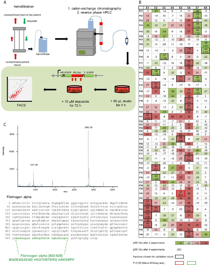
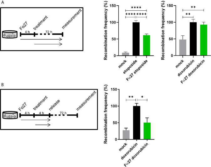
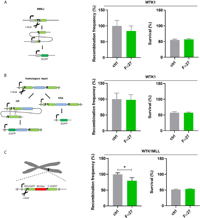
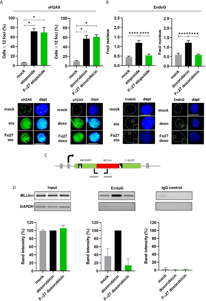
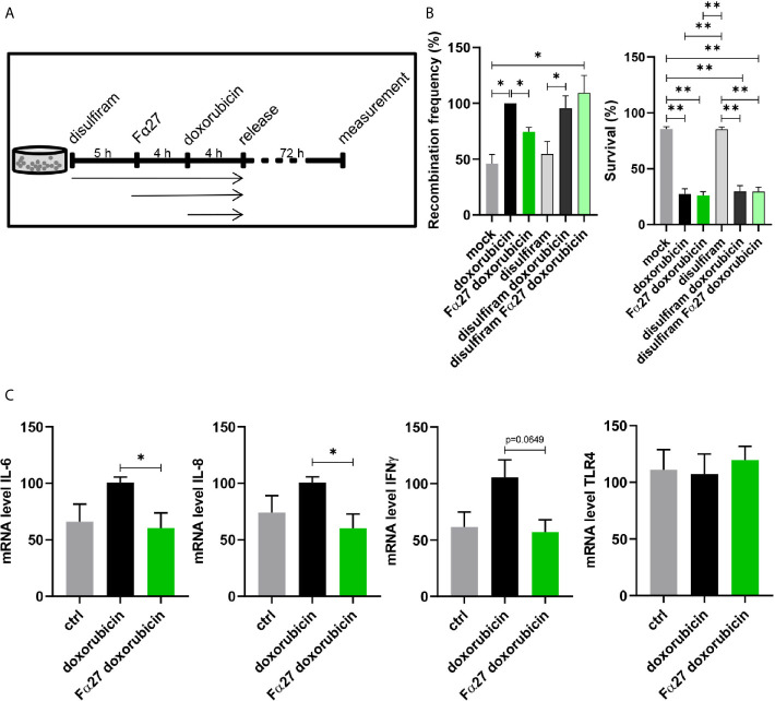
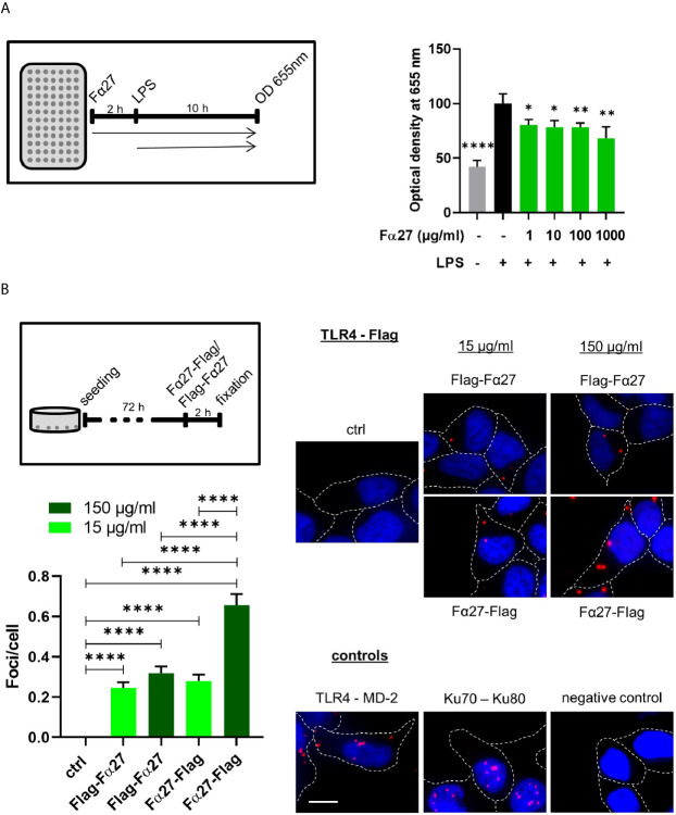
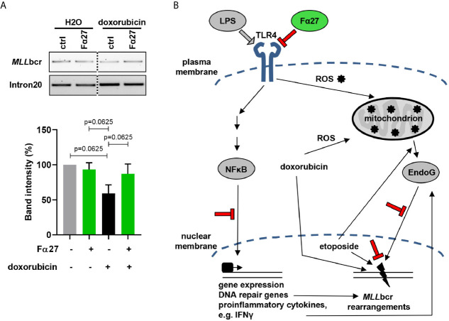
Rearrangements in the Mixed Lineage Leukemia breakpoint cluster region (MLLbcr) are frequently involved in therapy-induced leukemia, a severe side effect of anti-cancer therapies. Previous work unraveled Endonuclease G as the critical nuclease causing initial breakage in the MLLbcr in response to different types of chemotherapeutic treatment. To identify peptides protecting against therapy-induced leukemia, we screened a hemofiltrate-derived peptide library by use of an enhanced green fluorescent protein (EGFP)-based chromosomal reporter of MLLbcr rearrangements. Chromatographic purification of one active fraction and subsequent mass spectrometry allowed to isolate a C-terminal 27-mer of fibrinogen α encompassing amino acids 603 to 629. The chemically synthesized peptide, termed Fα27, inhibited MLLbcr rearrangements in immortalized hematopoietic cells following treatment with the cytostatics etoposide or doxorubicin. We also provide evidence for protection of primary human hematopoietic stem and progenitor cells from therapy-induced MLLbcr breakage. Of note, fibrinogen has been described to activate toll-like receptor 4 (TLR4). Dissecting the Fα27 mode-of action revealed association of the peptide with TLR4 in an antagonistic fashion affecting downstream NFκB signaling and pro-inflammatory cytokine production. In conclusion, we identified a hemofiltrate-derived peptide inhibitor of the genome destabilizing events causing secondary leukemia in patients undergoing chemotherapy.

Keywords: Endonuclease G; bioactive peptide; doxorubicin; hematopoietic stem and progenitor cells; inflammatory signaling; mixed lineage leukemia.


Conflict of interest statement

The authors declare that the research was conducted in the absence of any commercial or financial relationships that could be construed as a potential conflict of interest.

Figures

Figure 1
Figure 1
Identification of a human peptide from hemofiltrate reducing etoposide-induced rearrangements at the MLLbcr. (A) Principle of the peptide screen. Hemofiltrate was subjected to cation exchange chromatography and subsequent reversed phase high-performance liquid chromatography (HPLC) resulting in 384 eluate fractions, of which an aliquot of 90 µl each was added to a 2 ml culture of the cell line K562MLL with chromosomally integrated EGFP-based reporter construct for MLLbcr rearrangements. After a pre-treatment for 4 h the chemotherapeutic drug etoposide (10 µM) was included for 72 h. EGFP gene reconstitution was triggered by etoposide-induced MLLbcr breakage and recombination between two differentially mutated EGFP genes adjacent to the MLLbcr. MLLbcr rearrangements were thus measured as recombination frequencies determining the fraction of green fluorescent cells compared to the total population of live cells (FSC/SSC gate) by FACS analysis using the diagonal FL1-FL2 Dot Plot gate. (B) First round screening pattern. Relative changes of etoposide-induced recombination with versus without preincubation of cells with eluate are indicated for each HPLC fraction in %. Statistical significances of differences between mean recombination frequencies from two independent experiments according to χ2 test are visualized by gradual color changes, whereby red indicates increased and green decreased etoposide-induced recombination. For fractions with mean difference (Δ) ≥33% (p< 0.05 or SD<Δ) validation experiments were performed (black frames) resulting in n=4 (red or green frame: p<0.05 according to Mann-Whitney U test). (C) Identification of Fibrinogen alpha [603-629] by mass spectrometry (MALDI-MS) and Edman analysis of fraction E8F08 after HPLC purification. The MALDI-MS spectrum shows the single and the double charged peptide with a molecular weight of 2862 Da representing a C-terminal portion of fibrinogen α. See also Figure S1 .
Figure 2
Figure 2
Fα27 differentially affects MLLbcr rearrangements in WTK1MLL cells after etoposide and doxorubicin treatment. (A) Continuous exposure of WTK1MLL cells to peptide and chemotherapeutic drugs. WTK1MLL cells were treated for 4 h with 1 mg/ml Fα27 followed by 72 h with Fα27 plus 10 µM etoposide or 10 µg/ml Fα27 followed by Fα27 plus 0.5 µM doxorubicin, respectively (n=12 obtained in four independent experiments). EGFP-positive cells were counted after 72 h by FACS. After etoposide but not doxorubicin treatment Fα27 reduced the recombination frequency significantly. (B) Release of WTK1MLL cells after peptide- and doxorubicin treatment. WTK1MLL cells were treated for 4 h with 10 µg/ml Fα27 followed by 4 h with Fα27 plus 0.5 µM doxorubicin treatment (n=6 from two individual experiments). Thereafter, the cells were released by washing with PBS and seeding in fresh medium. FACS analysis showed a significant reduction of the doxorubicin-induced recombination frequency by Fα27 pre-treatment. Mean recombination frequencies induced by etoposide (8.5x10⁻⁵) and by doxorubicin (1.8x10⁻⁵) were set to 100% for each experimental day. All values are represented as mean +SEM (*p < 0.05, **p < 0.01, ****p < 0.0001).
Figure 3
Figure 3
Influence of Fα27 on I-SceI-induced MMEJ and homologous repair. (A) Scheme and results of MMEJ analysis following repair substrate cleavage by I-SceI. WTK1 cells were pre-treated with 1 mg/ml Fα27 for 4 h and then transfected with two plasmids, namely the repair construct for MMEJ and the I-SceI endonuclease expression plasmid (18). Targeted cleavage by I-SceI splits the mutated EJ-EGFP gene, so that MMEJ repair of the reporter permits EGFP reconstitution via pairing of adjacent microhomologies. EGFP-positive cells were measured by FACS 48 h post-transfection (n=9 obtained in three individual experiments). Mean recombination frequencies of controls were set to 100% each (absolute mean: 2.9x10⁻⁵). (B) Scheme and results of homologous repair analysis following cleavage by I-SceI. After cleavage by I-SceI either HR or SSA can reconstitute an intact EGFP gene. A 4 h pre-treatment with 1 mg/ml Fα27 and transfection with homologous repair reporter as well as I-SceI expression plasmid was followed by 48 h cultivation and FACS analysis (n=8-9 from three individual experiments). Mean recombination frequencies of controls were set to 100% each (absolute mean: 7.1x10⁻⁴). (C) Repair of I-SceI-induced DSBs in the same chromosomally integrated construct also used for detection of drug-induced MLLbcr rearrangements. WTK1MLL cells were treated with 1 mg/ml Fα27 for 4h, transfected with I-SceI expression plasmid and FACS analysis performed 48 h post-transfection (n=8-9 obtained in three independent experiments). Mean recombination frequencies of controls were set to 100% each (absolute mean: 1.2x10⁻⁴). All data represent mean +SEM (*p < 0.05). Corresponding survival data obtained by FACS analysis of live cells in the FSC/SSC plot are displayed in the right panel each.
Figure 4
Figure 4
Fα27 influences EndoG accumulation in nuclear foci during treatment and reduces local EndoG binding to the MLLbcr. (A) No effect of Fα27 on nuclear DNA damage level during chemotherapeutic treatment. WTK1 cells were treated for 4 h with 1 mg/ml Fα27 followed by inclusion of 10 µM etoposide for 4 h or 10 µg/ml Fα27 for 4 h followed by inclusion of 0.5 µM doxorubicin for 4 h. Fixed cells showed an increase of cells with ≥12 γH2AX foci/nucleus after etoposide or doxorubicin treatment but no decrease with Fα27 pre-treatment (n=4 obtained in two independent experiments). (B) Decrease of nuclear EndoG signals by Fα27 treatment. WTK1 cells were treated as described in (A) and immunofluorescently labeled for EndoG detection. The nuclear EndoG signals increased after etoposide (n=294-463 obtained in two independent experiments) or doxorubicin (n=296-485 obtained in two independent experiments) treatments and Fα27 significantly decreased the nuclear EndoG level. Insets display the highlighted region at two-fold magnification. Scale bars indicate 10 µM. White dashed lines encircle the DAPI-stained nucleus. All values represent mean +SEM (*p < 0.05; ****p < 0.0001). (C) Scheme of the MLLbcr region flanked by two mutated EGFP genes. The scheme visualizes the positions of the primers used for PCR after chromatin immunoprecipitation (ChIP). To analyze the binding of EndoG to the MLLbcr a ChIP was performed. Sonified DNA bound by proteins was immunoprecipitated with EndoG antibody or incubated with a control antibody. After DNA isolation a PCR was performed and band intensities evaluated. (D) Decreased EndoG binding to the MLLbcr after Fα27 treatment. WTK1MLL cells were treated for 4 h with 10 µg/ml Fα27 and additionally with 0.5 µM doxorubicin for 4 h. Two independent experiments for EndoG-ChIP and IgG-ChIP were performed. After PCR amplification of the MLLbcr in the recombination reporter, band intensities for input DNA and EndoG-ChIP samples after doxorubicin treatment were set to 100% and relative intensities calculated for the other samples including IgG-ChIP detected on the same membrane and with the same exposure time. EndoG binding to MLLbcr was increased after doxorubicin treatment and decreased with Fα27 treatment. Neither the input nor the control showed such a pattern. Control GAPDH PCR was positive for input but negative for ChIP samples. Data are shown as mean +SD. See also Figure S3 .
Figure 5
Figure 5
Visualization of Fα27 localization in HeLa cells. (A) Wide field image of HeLa cells at 60x magnification to observe cellular outlines and (B) image of the same field of view with spinning disc confocal fluorescence microscopy to visualize the localization of TAMRA-Fα27 present at 0.27 µM in the medium. Cells not taking up TAMRA-Fα27 appear dark within the bright background of medium containing TAMRA-Fα27. (C) Wide field image of a HeLa cell at 100x magnification to observe the cellular outline and (D) image of the same field of view with HILO fluorescence microscopy with single molecule sensitivity to visualize TAMRA-Fα27 present at 2.7 nM in the medium. The white dashed line indicates the cell membrane revealed in (C). The white arrow indicates a single TAMRA-Fα27 molecule visible as bright diffraction-limited spot. Bright intensity in the medium [upper left corner in (D)] comes from fast diffusing TAMRA-Fα27 molecules, whose signal is spread out over a larger area during the camera exposure time. Inset: mean intensity vs time plot of a small area encircling the spot indicated by the arrow. The signal of TAMRA-Fα27 is visible between ~0.05s and ~0.1s. Scale bar is 10 μm in (A, B) and 5 μm in (C, D).
Figure 6
Figure 6
Involvement of NFκB and TLR4 in Fα27 signaling. (A) Scheme for doxorubicin treatment protocol following exposure to the NFκB inhibitor disulfiram. WTK1MLL cells were treated for 5 h with 4 µM disulfiram followed by inclusion of 10 µg/ml Fα27 for 4 h and 0.5 µM doxorubicin for another 4 h. Then the cells were released by washing with PBS and seeded in fresh medium. (B) Influence of disulfiram under Fα27 and doxorubicin treatment. The recombination frequency measured by FACS showed a decrease of doxorubicin-induced MLLbcr rearrangements by Fα27 treatment (six independent experiments). This effect was lost in the presence of disulfiram. Mean recombination frequencies of doxorubicin treated cells were set to 100% each (absolute mean: 1.6x10⁻⁵). The panel in the right shows that survival was affected by doxorubicin but not by disulfiram. Data are represented as mean +SEM (*p < 0.05; **p < 0.01). (C) qRT-PCR of TLR4 downstream genes. The cells were treated with 10 µg/ml Fα27 for 4 h, 0.5 µM doxorubicin included for 4 h, total RNA isolated and mRNA subjected to qRT-PCR. Doxorubicin treatment increased mRNA expression of IL-6, IL-8 and IFNγ, whereas Fα27 reduced these doxorubicin-induced levels (n=5-6 obtained in three independent experiments). TLR4 mRNA expression was not changed. Statistical significances were calculated for doxorubicin-treated cells using Mann-Whitney-U test (*p < 0.05) and values are represented as mean +SEM. See also Figures S4 and S5 .
Figure 7
Figure 7
Fα27 binds TLR4 and reduces LPS stimulation. (A) Fα27 inhibits TLR4 activation by LPS. Scheme of experimental analysis of TLR4 signaling and results obtained in HEK-Blue hTLR4 cells pre-treated for 2 h with different concentrations of Fα27 followed by additional treatment with 10 ng/ml LPS for 10 h. TLR4 stimulation led to a release of secreted embryonic alkaline phosphatase (SEAP) detected at OD 655 nm. LPS treatment increased SEAP release which was reduced by Fα27 pre-treatment. Mean OD values of LPS-treated controls without peptide were set to 100% in each experiment. Data (n=12 obtained in two experiments) are shown as mean +SEM and statistics were calculated relative to LPS stimulation without peptide (*p < 0.05; **p < 0.01; ****p < 0.0001). (B) TLR4 and Fα27 interaction. HEK-Blue hTLR4 cells were seeded 72 h before the experiment and treated for 2 h with N-terminally tagged Flag-Fα27, C-terminally tagged Fα27-Flag or water (ctrl). PLA analysis showed an interaction between both tagged Fα27 peptides and the TLR4. Two positive controls are shown: PLA for TLR4 and its binding partner MD-2 on the cellular surface and for Ku70-Ku80 complexes in the nucleus. The negative control was done with PLA substrates but without antibodies. Margins of cells are marked by white stippled lines and the scale bar indicates 10 µM (n=187-425 from two independent experiments each; mean +SEM; ****p < 0.0001). See also Figure S6 .
Figure 8
Figure 8
Fα27 prevents MLLbcr breakage in HSPCs. (A) MLLbcr breakage analysis after doxorubicin and Fα27 treatment of HSPCs. HSPCs were treated 4 h with 10 µg/ml Fα27 followed by 4 h of treatment with Fα27 plus 0.072 µM doxorubicin. After genomic DNA isolation breakage was assessed by PCR targeting the endogenous MLLbcr locus. Doxorubicin treatment increased breakage as indicated by a 40% decrease of the band intensity, which was reduced in Fα27 pre-treated cells by only 13%. Data from five independent experiments are shown as mean +SEM (Friedman-Test indicating p=0.0087, followed by Wilcoxon test). The framed image shows MLLbcr PCR amplifications from the same gel exposed with the same intensity and cropped at the dotted line. MLL intron20 was amplified as control. (B) Model of MLLbcr protection by Fα27. LPS activation of TLR4 leads to NFκB-mediated gene expression of DNA repair genes and proinflammatory cytokines like type I interferons (51). The interferon IFNγ can increase EndoG release from the mitochondria (52). Etoposide, a topoisomerase II inhibitor, induces DNA damage and EndoG release from the mitochondria (53). Doxorubicin, a drug with a broad spectrum of cell damage, intercalates into the DNA, which promotes translocation of EndoG from mitochondria to the nucleus. Nuclear EndoG cleaves the MLLbcr predicted to form pronounced secondary structures. Stimulation of TLR4, such as by LPS, was as well reported to increase ROS production in mitochondria (54), again inducing DNA damage. We show that Fα27 blocks TLR4 activation and downstream NFκB signaling. Furthermore, EndoG accumulation in the nucleus is reduced and the MLL gene protected from breakage. Findings made in this work are indicated in red color.

Similar articles

Cited by

References

    1. De Braekeleer M, Morel F, Le Bris MJ, Herry A, Douet-Guilbert N. The MLL Gene and Translocations Involving Chromosomal Band 11q23 in Acute Leukemia. Anticancer Res (2005) 25:1931–44. - PubMed
    1. Gole B, Wiesmüller L. Leukemogenic Rearrangements at the Mixed Lineage Leukemia Gene (MLL)—Multiple Rather Than a Single Mechanism. Front Cell Dev Biol (2015) 3:41. 10.3389/fcell.2015.00041 - DOI - PMC - PubMed
    1. Meyer C, Burmeister T, Gröger D, Tsaur G, Fechina L, Renneville A, et al. . The MLL Recombinome of Acute Leukemias in 2017. Leukemia (2018) 32:273–84. 10.1038/leu.2017.213 - DOI - PMC - PubMed
    1. Liu H, Cheng EHY, Hsieh JJD. MLL Fusions: Pathways to Leukemia. Cancer Biol Ther (2009) 8:1204–11. 10.4161/cbt.8.13.8924 - DOI - PMC - PubMed
    1. Gleissner B, Goekbuget N, Rieder H, Arnold R, Schwartz S, Diedrich H, et al. . CD10- Pre-B Acute Lymphoblastic Leukemia (ALL) Is a Distinct High-Risk Subgroup of Adult ALL Associated With a High Frequency of MLL Aberrations: Results of the German Multicenter Trials for Adult All (GMALL). Blood (2005) 106:4054–6. 10.1182/blood-2005-05-1866 - DOI - PubMed