. 2021 Jun 18:11:686090.
doi: 10.3389/fcimb.2021.686090. eCollection 2021.

Characterisation of Bacteriophage-Encoded Depolymerases Selective for Key Klebsiella pneumoniae Capsular Exopolysaccharides

Affiliations

Characterisation of Bacteriophage-Encoded Depolymerases Selective for Key Klebsiella pneumoniae Capsular Exopolysaccharides

George Blundell-Hunter et al. Front Cell Infect Microbiol. .

Abstract

Capsular polysaccharides enable clinically important clones of Klebsiella pneumoniae to cause severe systemic infections in susceptible hosts. Phage-encoded capsule depolymerases have the potential to provide an alternative treatment paradigm in patients when multiple drug resistance has eroded the efficacy of conventional antibiotic chemotherapy. An investigation of 164 K. pneumoniae from intensive care patients in Thailand revealed a large number of distinct K types in low abundance but four (K2, K51, K1, K10) with a frequency of at least 5%. To identify depolymerases with the capacity to degrade capsules associated with these common K-types, 62 lytic phage were isolated from Thai hospital sewage water using K1, K2 and K51 isolates as hosts; phage plaques, without exception, displayed halos indicative of the presence of capsule-degrading enzymes. Phage genomes ranged in size from 41-348 kb with between 50 and 535 predicted coding sequences (CDSs). Using a custom phage protein database we were successful in applying annotation to 30 - 70% (mean = 58%) of these CDSs. The largest genomes, of so-called jumbo phage, carried multiple tRNAs as well as CRISPR repeat and spacer sequences. One of the smaller phage genomes was found to contain a putative Cas type 1E gene, indicating a history of host DNA acquisition in these obligate lytic phage. Whole-genome sequencing (WGS) indicated that some phage displayed an extended host range due to the presence of multiple depolymerase genes; in total, 42 candidate depolymerase genes were identified with up to eight in a single genome. Seven distinct virions were selected for further investigation on the basis of host range, phage morphology and WGS. Candidate genes for K1, K2 and K51 depolymerases were expressed and purified as his6-tagged soluble protein and enzymatic activity demonstrated against K. pneumoniae capsular polysaccharides by gel electrophoresis and Anton-Paar rolling ball viscometry. Depolymerases completely removed the capsule in K-type-specific fashion from K. pneumoniae cells. We conclude that broad-host range phage carry multiple enzymes, each with the capacity to degrade a single K-type, and any future use of these enzymes as therapeutic agents will require enzyme cocktails for utility against a range of K. pneumoniae infections.

Keywords: Klebsiella pneumoniae; alternative antibacterial therapy; bacteriophage; capsular polysaccharide; capsule depolymerase; jumbo phage; whole-genome sequencing (WGS).


Conflict of interest statement

The authors declare that the research was conducted in the absence of any commercial or financial relationships that could be construed as a potential conflict of interest.

Figures

Figure 1
Figure 1
Phylogeny of Thai K pneumoniae- selective phage. (A) MASH tree with degree of genetic similarity amongst the 28 phage genomes sequenced in this study. (B) Phylogenetic tree prepared using Archaeopteryx showing the relationship of the 28 phage genomes (in red) to 314 published Klebsiella phage genomes.
Figure 2
Figure 2
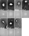
Representative transmission electron micrographs and plaque morphology of the seven exemplars for each of the genetically distinct phage groups defined in this study and shown in Table 2 . (A) GBH001 (mean capsid diameter 63.4 nm; mean tail length N/A; n = 10) (B) GBH014 (64.0 nm; N/A; n = 7) (C) GBH019 (132.7 nm; 163.4 nm; n = 9) (D) GBH029 (73.8 nm; 188.8 nm; n = 7) (E) GBH033 (86.7 nm; 105.2 nm; n = 8) (F) GBH038 (72.0 nm; N/A; n = 6) (G) GBH054 (81.2 nm; 256.4 nm; n = 11).
Figure 3
Figure 3
Circular genomic map of Jumbo phage GBH019. Genes involved in virion structure (orange), host interaction (green), nucleotide metabolism (pink), DNA replication and repair (blue), and DNA transcription (magenta) are shown as the positions of six tRNAs (red) and a putative CRISPR array (purple). %GC is shown in the centre of the figure. Hypothetical proteins are shown in cream. Figure generated using DNA Plotter release 18.1.0.
Figure 4
Figure 4
Degradation of high-molecular-weight capsular polysaccharide from K. pneumoniae SR65 (A), SR3 (B) and TU16 (C) following incubation at 37°C with the corresponding K1, K2 and K51 depolymerases. 10% SDS-PAGE gels; 5 μl Color Prestained Protein Standard, Broad Range (10-250 kDa) (New England Biolabs UK) as ladder; 22.5 μl loaded into each lane. Enzyme-mediated reductions in polymer viscosity as determined by Anton-Paar rolling ball viscometry are also shown alongside the corresponding gels. No polymer degradation was observed in the absence of enzyme. ****P = < 0.0001 (unpaired t-test with Welch’s correction between treated and untreated groups).
Figure 5
Figure 5
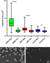
Box and whiskers plot of the impact on capsules of the target K. pneumoniae isolates SR65 (K1), SR3 (K2) and TU16 (K51) following 90 min incubation at 37°C with the corresponding K1, K2 and K51 depolymerases. SR65 Neg, negative control (PBS incubation), n = 319, mean capsule area μm2 3.41 ± 1.00 (Mean ± 1SD); SR65 Pos, K1 depolymerase-exposed, n = 74, 1.49 ± 0.34; SR3 Neg, n = 119, 1.96 ± 0.58; SR3 Pos, n = 66, 1.41 ± 0.42; TU16 Neg, n = 84, 1.54 ± 0.42; TU16 Pos, n = 170, 1.36 ± 0.31. ****P = < 0.0001, ***P = < 0.002 (unpaired t-test with Welch’s correction between treated and untreated groups). Also shown: phase contrast microscopy images of nigrosin staining for isolate SR65 (K1) after 90 min incubation in PBS (A) and with K1 depolymerase (B). Capsule can be seen in (A) as a bright halo around the cell.

References

    1. Adams M. H., Park B. H. (1956). An Enzyme Produced by a Phage Host-Cell System. II. The Properties of the Polysaccharide Depolymerase. Virology 2, 719–736. 10.1016/0042-6822(56)90054-x - DOI - PubMed
    1. Adriaenssens E., Brister J. R. (2017). How to Name and Classify Your Phage: An Informal Guide. Viruses 9, 70. 10.3390/v9040070 - DOI - PMC - PubMed
    1. Adriaenssens E. M., Sullivan M. B., Knezevic P., van Zyl L. J., Sarkar B. L., Dutilh B. E., et al. . (2020). Taxonomy of Prokaryotic Viruses: 2018-2019 Update From the ICTV Bacterial and Archaeal Viruses Subcommittee. Arch. Virol. 165, 1253–1260. 10.1007/s00705-020-04577-8 - DOI - PubMed
    1. Al-Shayeb B., Sachdeva R., Chen L. X., Ward F., Munk P., Devoto A., et al. . (2020). Clades of Huge Phages From Across Earth’s Ecosystems. Nature 578, 425–431. 10.1038/s41586-020-2007-4 - DOI - PMC - PubMed
    1. Avery O. T., Dubos R. (1930). The Specific Action of a Bacterial Enzyme on Pneumococci of Type III. Science 72, 151–152. 10.1126/science.72.1858.151 - DOI - PubMed

Publication types