Characterizing the Xenoma of Vairimorpha necatrix Provides Insights Into the Most Efficient Mode of Microsporidian Proliferation
- PMID: 34222053
- PMCID: PMC8242933
- DOI: 10.3389/fcimb.2021.699239
Characterizing the Xenoma of Vairimorpha necatrix Provides Insights Into the Most Efficient Mode of Microsporidian Proliferation
Abstract
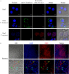
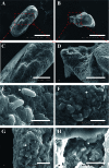
Microsporidia are a group of obligated intracellular parasites that can infect nearly all vertebrates and invertebrates, including humans and economic animals. Microsporidian Vairimorpha necatrix is a natural pathogen of multiple insects and can massively proliferate by making tumor-like xenoma in host tissue. However, little is known about the subcellular structures of this xenoma and the proliferation features of the pathogens inside. Here, we characterized the V. necatrix xenoma produced in muscle cells of silkworm midgut. In result, the whitish xenoma was initially observed on the 12th day post infection on the outer surface of the midgut and later became larger and numerous. The observation by scanning electronic microscopy showed that the xenoma is mostly elliptical and spindle with dense pathogen-containing protrusions and spores on the surface, which were likely shedding off the xenoma through exocytosis and could be an infection source of other tissues. Demonstrated with transmission electron microscopy and fluorescent staining, the xenoma was enveloped by a monolayer membrane, and full of vesicle structures, mitochondria, and endoplasmic reticulum around parasites in development, suggesting that high level of energy and nutrients were produced to support the massive proliferation of the parasites. Multiple hypertrophic nuclei were found in one single xenoma, indicating that the cyst was probably formed by fusion of multiple muscle cells. Observed by fluorescence in situ hybridization, pathogens in the xenoma were in merongony, sporogony, and octosporogony, and mature stages. And mature spores were pushed to the center while vegetative pathogens were in the surface layer of the xenoma. The V. necatrix meront usually contained two to three nuclei, and sporont contained two nuclei and was wrapped by a thick membrane with high electron density. The V. necatrix sporogony produces two types of spores, the ordinary dikaryotic spore and unicellular octospores, the latter of which were smaller in size and packed in a sporophorous vesicle. In summary, V. necatrix xenoma is a specialized cyst likely formed by fusion of multiple muscle cells and provides high concentration of energy and nutrients with increased number of mitochondria and endoplasmic reticulum for the massive proliferation of pathogens inside.
Keywords: Vairimorpha necatrix; microsporidia; proliferation; subcellular structure; xenoma.
Copyright © 2021 Li, Fang, He, Wang, Meng, Yu and Zhou.
Conflict of interest statement
The authors declare that the research was conducted in the absence of any commercial or financial relationships that could be construed as a potential conflict of interest. The reviewer JX declared a shared affiliation, with no collaboration, with one of the authors, ZZ, to the handling editor at the time of review.
Figures


References
-
- Azevedo C. (1987). Fine Structure of the Microsporidan Abelspora Portucalensis Gen.N., Sp.N. (Microsporida) Parasite of the Hepatopancreas of Carcinus Maenas (Crustacea, Decapoda). J. Invertebrate Pathol. 49 (1), 83–92. 10.1016/0022-2011(87)90129-7 - DOI
-
- Cali A., Kent M., Sanders J., Pau C., Takvorian P. M. (2012). Development, Ultrastructural Pathology, and Taxonomic Revision of the Microsporidial Genus, Pseudoloma and its Type Species Pseudoloma Neurophilia, in Skeletal Muscle and Nervous Tissue of Experimentally Infected Zebrafish Danio Rerio . J. Eukaryotic Microbiol. 59 (1), 40–48. 10.1111/j.1550-7408.2011.00591.x - DOI - PMC - PubMed
-
- Cali A., Takvorian P. M. (1999). “Developmental Morphology and Life Cycles of the Microsporidia” in The Microsporidia and Microsporidiosis. (WILEY Blackwell; ), 85–128.
Publication types
MeSH terms
Supplementary concepts
LinkOut - more resources
Full Text Sources

