

Benfotiamine reduced collagen IV contents of sciatic nerve in hyperglycemic rats
- PMID: 34222057
- PMCID: PMC8212243
- DOI: 10.1007/s40200-020-00666-2
Benfotiamine reduced collagen IV contents of sciatic nerve in hyperglycemic rats
Abstract
Background: Neuropathy as a common complication of hyperglycemia in diabetic patients is probably caused by metabolic and structural changes in extracellular matrix (ECM) of peripheral nerves. This study was designed to evaluate the effects of benfotiamine (BT) on the structural, biological and mechanical characteristics of rat sciatic nerve in hyperglycemic condition.
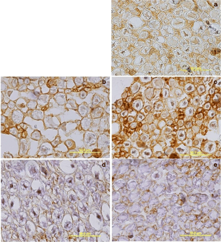
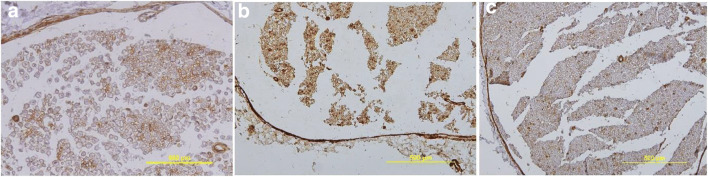
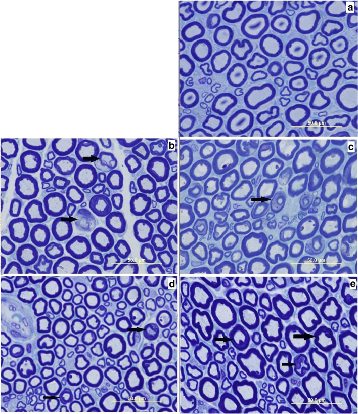
Materials and methods: Forty eight adult male Wistar rats were assigned to 6 groups (n = 8): control (healthy rats with no treatment; C), positive control (healthy rats received BT treatment; B), negative control groups 1&2 (hyperglycemic rats kept for 4 and/or 8 weeks; 4WD and 8WD, respectively) and experimental groups 1&2 (hyperglycemic rats treated by daily oral gavage of 100 mg kg- 1 body weight BT for 4 and/or 8 weeks; 4WD + BT and 8WD + BT, respectively). Hyperglycemia was induced by a single intraperitoneal injection of of streptozotocin (55 mg kg- 1 body weight). After a period of experimental period (4 and/or 8 weeks) rats were sacrificed and from each two segments (1 cm length) of left sciatic nerve were sampled. These samples were prepared for histological examinations (light and electron microscopy), collagen IV immunohistochemistry and strength tensile test.
Results: In comparison to control groups, in 4WD and 8WD groups the amount of type IV collagen was increased, the structure of myelin sheath and nerve fibers were extensively altered and the tensile strength was significantly decreased (p < 0.05) while in 4WD + BT and 8WD + BT groups these abnormalities were attenuated.
Conclusions: It seems that BT treatment may rescue the sciatic nerve from the hyperglycemic-induced ECM structural abnormality. This beneficial advantage of BT is likely exerted through the modification of glucose metabolism pathways.
Keywords: Benfotiamine; Collagen IV; Diabetes; Sciatic nerve; Tensile strength.
© Springer Nature Switzerland AG 2020.
Conflict of interest statement
Conflict of interestThe authors declare no conflict of interest.
Figures


References
-
- Rassouli MB, Ghayour MB, Ghayour N. Microvascular complications of diabetes. J Biol Sci. 2010;10:411–23.
-
- Diabéticas Periféricas N, et al. Diabetic peripheral neuropathies: a morphometric overview. Int J Morphol. 2010;28(1):51–64.
-
- Vinik AI, Nevoret M, Casellini C, Parson H. Diabetic neuropathy. Endocrinol Metab Clin North Am. 2013;42(4):747-87 (Elsevier Inc). - PubMed
-
- Bruschi LKM, et al. Diabetes mellitus and diabetic peripheral neuropathy. Open J Endocr Metab Dis. 2017;07(01):12–21.
LinkOut - more resources
Full Text Sources
