. 2021 Jun 16:9:681501.
doi: 10.3389/fbioe.2021.681501. eCollection 2021.

High-Efficient Production of Adipose-Derived Stem Cell (ADSC) Secretome Through Maturation Process and Its Non-scarring Wound Healing Applications

Affiliations

High-Efficient Production of Adipose-Derived Stem Cell (ADSC) Secretome Through Maturation Process and Its Non-scarring Wound Healing Applications

Young-Hyeon An et al. Front Bioeng Biotechnol. .

Abstract

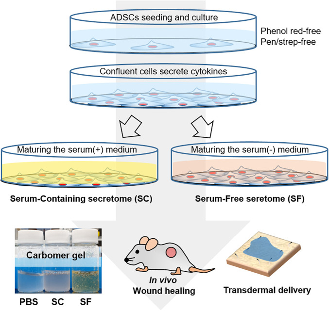
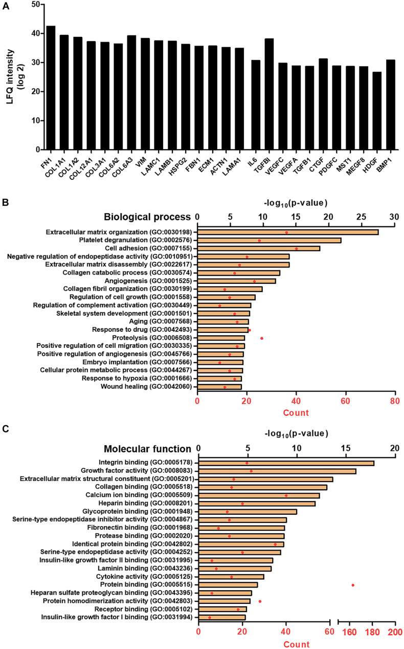
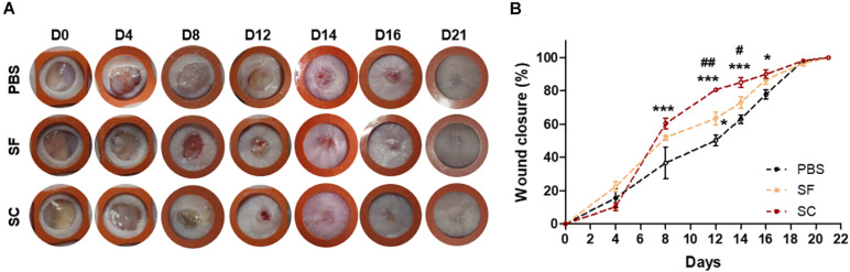
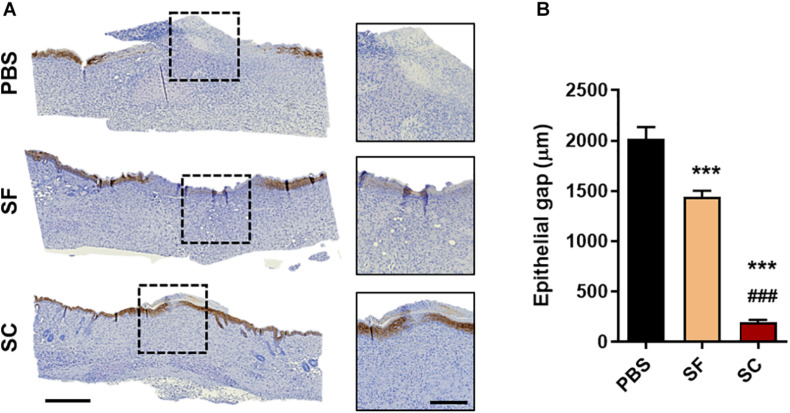
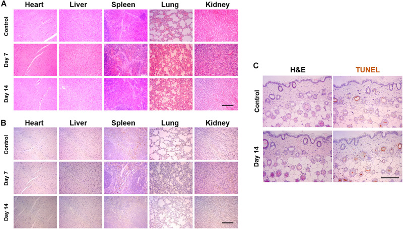
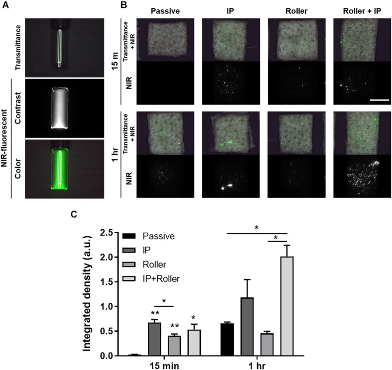
Recently, the stem cell-derived secretome, which is the set of proteins expressed by stem cells and secreted into the extracellular space, has been demonstrated as a critical contributor for tissue repair. In this study, we have produced two sets of high concentration secretomes from adipose-derived mesenchymal stem cells (ADSCs) that contain bovine serum or free of exogenous molecules. Through proteomic analysis, we elucidated that proteins related to extracellular matrix organization and growth factor-related proteins are highly secreted by ADSCs. Additionally, the application of ADSC secretome to full skin defect showed accelerated wound closure, enhanced angiogenic response, and complete regeneration of epithelial gaps. Furthermore, the ADSC secretome was capable of reducing scar formation. Finally, we show high-dose injection of ADSC secretome via intraperitoneal or transdermal delivery demonstrated no detectable pathological conditions in various tissues/organs, which supports the notion that ADSC secretome can be safely utilized for tissue repair and regeneration.

Keywords: proteomic analysis; secretome; skin regeneration; stem cells; tissue repair.


Conflict of interest statement

DHK, EL, JWK, and S-DK were employed by the company, Senior Science & Life, Inc. The remaining authors declare that the research was conducted in the absence of any commercial or financial relationships that could be construed as a potential conflict of interest.

Figures

SCHEME 1
SCHEME 1
The schematic illustration of the experimental procedure. The serum-containing (SC) and serum-free secretome (SF) were harvested by maturing the fully confluent adipose tissue-derived stem cells (ADSCs). The secretome was applied to the in vivo skin wound and tried to be delivered transdermally using the ex vivo porcine skin.
FIGURE 1
FIGURE 1
Proteomic analysis of SF. (A) LFQ intensity of representative extracellular matrix protein and growth factors, measured by LC/MS-MS. Gene ontology (GO) analysis representing the top 20 GO terms and its number of enrichment in (B) biological process and (C) molecular function.
FIGURE 2
FIGURE 2
The ADSC secretome accelerated wound closure. (A) Photographs of the wound (rubber ring diameter = 9 mm). (B) Wound closure profiles by measuring the wound size (*compared with the PBS group; #compared with the SF group;*#p < 0.05; ##p < 0.01; ***p < 0.001).
FIGURE 3
FIGURE 3
Immunohistochemical staining of cytokeratin-10 on day 14. (A) Representative microscopy images (Scale bar = 500 μm in low magnified images and 200 μm in high magnified images) and (B) the gap between the regenerated epithelial cells (***###p < 0.001).
FIGURE 4
FIGURE 4
Hematoxylin and eosin (H&E) staining of the wound on days 14 and 21. (A) Light microscopy images of the wound bed representing the panniculus adiposus (black arrow) and carnosus (red arrow) (Scale bar = 500 μm). (B) Quantitative measurement of the panniculus gap at days 14 and 21 (*compared with the PBS group; #compared with the SF group; *p < 0.05; **##p < 0.01; ***###p < 0.001).
FIGURE 5
FIGURE 5
Masson’s trichrome (MTC) staining of the wounds at day 21. (A) Representative light microscopy images (Scale bar = 100 μm). The qualitative analysis of the skin regeneration (B) collagen deposition, (C) extracellular matrix (ECM) fiber alignment, and (D) the number of skin appendages (*compared with the PBS group; #compared with the SF group; p < 0.05; **p < 0.01; ***p < 0.001).
FIGURE 6
FIGURE 6
In vivo angiogenesis evaluation with the immunohistochemical staining (IHC) of alpha-smooth muscle actin (α-SMA). (A) Representative light microscopy images at regenerated wounds (Scale bar = 100 μm). (B) The number of newly formed vessels in the high-power field (HPF) magnification images (n = 10–15) (*compared with the PBS group; #compared with the SF group; *,#p < 0.05; ***p < 0.001).
FIGURE 7
FIGURE 7
Pathological analysis of SF via intraperitoneal injection and applying to the skin surface. The 50 μl of SF was injected every other day until 2 weeks or applied to the dorsal skin surface of mice. (A) Hematoxyline and eosin (H&E) and (B) TUNEL staining of the tissues harvested from the heart, the liver, the spleen, the lung, and the kidney. (C) H&E- and TUNEL-stained images of the skin, which did not display any toxical responses (Scale bar = 200 μm).
FIGURE 8
FIGURE 8
Ex vivo transdermal delivery of NIR dye-labeled SF. (A) The NIR dye, ZW800-1C-NHS-ester, was conjugated with proteins in the secretome. (B) Macroscopic visualization of transport of secretome into the ex vivo porcine skin with the assistance of physical penetration enhancers (PPEs). (C) The quantification data based on the FLARE images indicated the combinatorial application of IP and roller enhanced the transport of secretome transdermally (*p < 0.05; **p < 0.01) (Scale bar = 1 cm).

References

    1. Alegre M. L. (2020). The anti-inflammatory function of the apoptotic secretome. Am. J. Transplant. 20 1471–1471. 10.1111/ajt.15990 - DOI - PubMed
    1. An Y. H., Park M. J., Lee J., Ko J., Kim S. H., Kang D. H., et al. (2020). Recent advances in the transdermal delivery of protein therapeutics with a combinatorial system of chemical adjuvants and physical penetration enhancements. Adv. Ther. Germany 3:1900116. 10.1002/adtp.201900116 - DOI
    1. Bai H. T., Kyu-Cheol N., Wang Z. H., Cui Y. T., Liu H., Liu H., et al. (2020). Regulation of inflammatory microenvironment using a self-healing hydrogel loaded with BM-MSCs for advanced wound healing in rat diabetic foot ulcers. J. Tissue Eng. 11:2041731420947242. - PMC - PubMed
    1. Balaji S., King A., Marsh E., LeSaint M., Bhattacharya S. S., Han N., et al. (2015). The role of interleukin-10 and hyaluronan in murine fetal fibroblast function in vitro: implications for recapitulating fetal regenerative wound healing. PLoS One 7:10. - PMC - PubMed
    1. Bari E., Perteghella S., Di Silvestre D., Sorlini M., Catenacci L., Sorrenti M., et al. (2018). Pilot production of mesenchymal stem/stromal freeze-dried secretome for cell-free regenerative nanomedicine: a validated GMP-compliant process. Cells. 7:190. 10.3390/cells7110190 - DOI - PMC - PubMed