Review
. 2021 Jun 18:8:664404.
doi: 10.3389/fsurg.2021.664404. eCollection 2021.

Current Applications and Future Directions of Bioengineering Approaches for Bladder Augmentation and Reconstruction

Affiliations
Review

Current Applications and Future Directions of Bioengineering Approaches for Bladder Augmentation and Reconstruction

Xuesheng Wang et al. Front Surg. .

Abstract

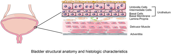
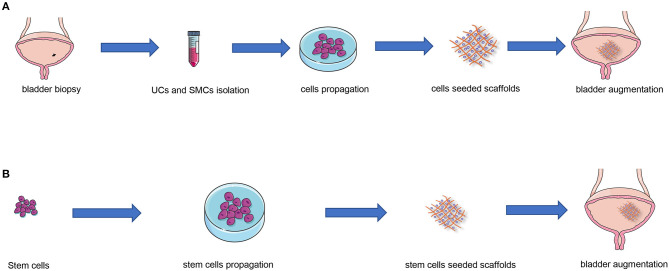
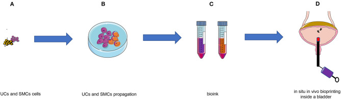
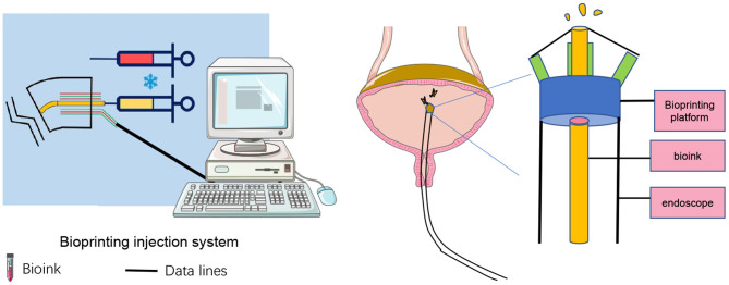
End-stage neurogenic bladder usually results in the insufficiency of upper urinary tract, requiring bladder augmentation with intestinal tissue. To avoid complications of augmentation cystoplasty, tissue-engineering technique could offer a new approach to bladder reconstruction. This work reviews the current state of bioengineering progress and barriers in bladder augmentation or reconstruction and proposes an innovative method to address the obstacles of bladder augmentation. The ideal tissue-engineered bladder has the characteristics of high biocompatibility, compliance, and specialized urothelium to protect the upper urinary tract and prevent extravasation of urine. Despite that many reports have demonstrated that bioengineered bladder possessed a similar structure to native bladder, few large animal experiments, and clinical applications have been performed successfully. The lack of satisfactory outcomes over the past decades may have become an important factor hindering the development in this field. More studies should be warranted to promote the use of tissue-engineered bladders in clinical practice.

Keywords: 3D bioprinting; bladder augmentation; bladder reconstruction; scaffolds; tissue engineering.


Conflict of interest statement

The authors declare that the research was conducted in the absence of any commercial or financial relationships that could be construed as a potential conflict of interest. The reviewer PZ declared a shared affiliation, with no collaboration, with the authors to the handling editor at the time of the review.

Figures

Figure 1
Figure 1
Bladder structural anatomy and histologic characteristics. The bladder walls consist of four layers: urothelium; lamina propria; muscular layer and serosal layer. The urothelium, composed of umbrella cells, intermediate cells, basal cells, basal membrane lines the bladder lumen and forms the urine-body barrier. The lamina propria is a connective tissue layer that contains nerves and vessels. The detrusor muscle layer consisted of longitudinal and transverse muscles that provides structural support to the bladder and facilitates its physiological functions of filling and emptying. The serosal layer covering the external surface is the outermost layer.
Figure 2
Figure 2
Tissue engineering strategies for autologous urothelial cells and stem cells. (A) UCs and SMCs obtained from the biopsy material first proliferate in the cell culture incubator; then the proliferated cells are subsequently reseeded into a tissue-engineered scaffold; eventually, the scaffold with cells was reimplanted into the same host. (B) Firstly, stem cells proliferate in the cell culture incubator; subsequently, the proliferated cells are seeded into a tissue-engineered scaffold; eventually, the scaffold with cells was implanted into the host. UCs, urothelial cells; SMCs, smooth muscle cells.
Figure 3
Figure 3
Schematic of in situ in vivo bioprinting taking the case of bladder augmentation. (A) UCs and SMCs were harvested from biopsy material; (B) UCs and SMCs proliferated in vitro; (C) Bioinks containing UCs and SMCs; (D) in situ in vivo bioprinting after bladder augmentation with biomaterial. UCs, urothelial cells; SMCs, smooth muscle cells.
Figure 4
Figure 4
Schematic of in situ in vivo bioprinting inside a bladder.

Similar articles

Cited by

References

    1. Chua ME, Farhat WA, Ming JM, McCammon KA. Review of clinical experience on biomaterials and tissue engineering of urinary bladder. World J Urol. (2020) 38:2081–93. 10.1007/s00345-019-02833-4 - DOI - PubMed
    1. Biers SM, Venn SN, Greenwell TJ. The past, present and future of augmentation cystoplasty. BJU Int. (2012) 109:1280–93. 10.1111/j.1464-410X.2011.10650.x - DOI - PubMed
    1. Chen J-L, Kuo H-C. Long-term outcomes of augmentation enterocystoplasty with an ileal segment in patients with spinal cord injury. J Formos Med Assoc. (2009) 108:475–80. 10.1016/S0929-6646(09)60095-4 - DOI - PubMed
    1. Gilbert SM, Hensle TW. Metabolic consequences and long-term complications of enterocystoplasty in children: a review. J Urol. (2005) 173:1080–6. 10.1097/01.ju.0000155248.57049.4e - DOI - PubMed
    1. Atala A, Bauer SB, Soker S, Yoo JJ, Retik AB. Tissue-engineered autologous bladders for patients needing cystoplasty. Lancet. (2006) 367:1241–6. 10.1016/S0140-6736(06)68438-9 - DOI - PubMed