. 2021 Mar 12;3(2):fcab035.
doi: 10.1093/braincomms/fcab035. eCollection 2021.

Evaluating cognitive profiles of patients undergoing clinical amyloid-PET imaging

Affiliations

Evaluating cognitive profiles of patients undergoing clinical amyloid-PET imaging

Flavia Loreto et al. Brain Commun. .

Abstract

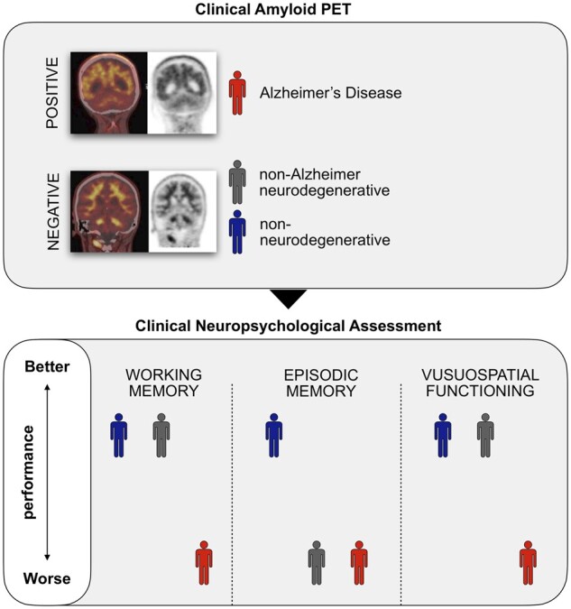
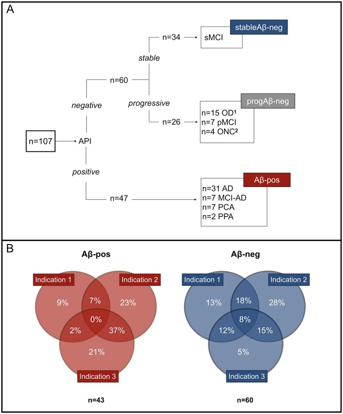
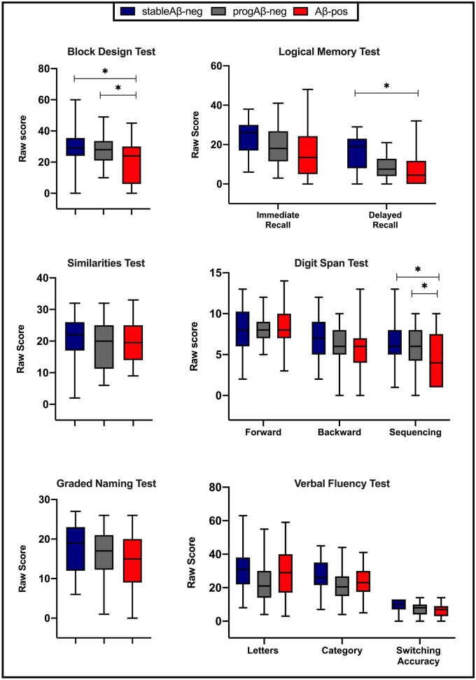
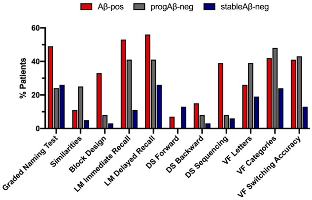
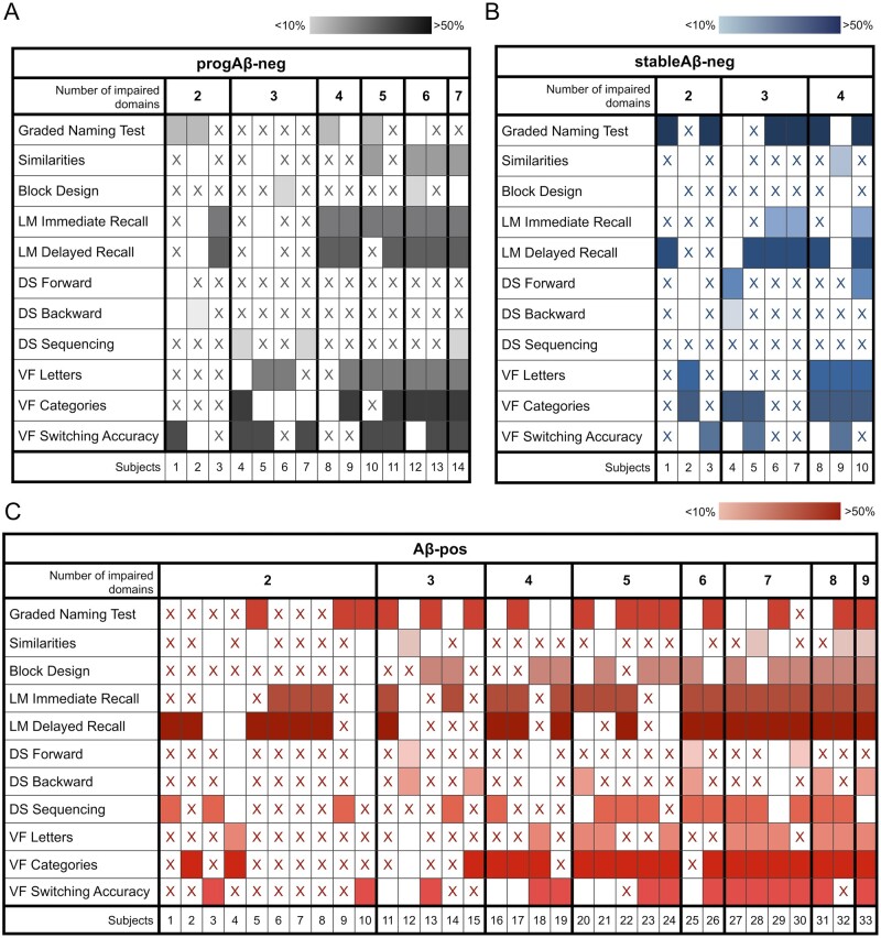
Episodic memory impairment and brain amyloid-beta are two of the main hallmarks of Alzheimer's Disease. In the clinical setting, these are often evaluated through neuropsychological testing and amyloid PET imaging, respectively. The use of amyloid PET in clinical practice is only indicated in patients with substantial diagnostic uncertainty due to atypical clinical presentation, multiple comorbidities and/or early age of onset. The relationship between amyloid-beta and cognition has been previously investigated, but no study has examined how neuropsychological features relate to the presence of amyloid pathology in the clinical population that meets the appropriate use criteria for amyloid PET imaging. In this study, we evaluated a clinical cohort of patients (n = 107) who presented at the Imperial Memory Clinic and were referred for clinical amyloid PET and neuropsychological assessment as part of their diagnostic workup. We compared the cognitive performance of amyloid-positive patients (Aβ-pos, n = 47) with that of stable amyloid-negative (stableAβ-neg, n = 26) and progressive amyloid-negative (progAβ-neg, n = 34) patients. The amyloid-positive group performed significantly worse than both amyloid-negative groups in the visuospatial and working memory domains. Episodic memory performance, however, effectively differentiated the amyloid-positive group from the stable but not the progressive amyloid-negative group. On affective questionnaires, the stable amyloid-negative group reported significantly higher levels of depression than the amyloid-positive group. In our clinical cohort, visuospatial dysfunction and working memory impairment were better indicators of amyloid positivity than episodic memory dysfunction. These findings highlight the limited value of isolated cognitive scores in patients with atypical clinical presentation, comorbidities and/or early age of onset.

Keywords: Alzheimer’s disease; amyloid PET imaging; cognitive testing; dementia; neuropsychology.


Figures

Graphical Abstract
Graphical Abstract
Figure 1
Figure 1
Group definition flowchart and Appropriate Use Criteria classification. (A) This figure illustrates the group characterization and sample sizes. The amyloid-negative (Aβ-neg) group was divided into progressive (progAβ-neg) and stable (stableAβ-neg) based on the presence or absence of clinical progression and/or concomitant neurological condition. Further details on the progAβ-neg group are as follows. 1OD (n = 15): FTD (n = 6), PSP (n = 1), CBS (n = 1), primary progressive MS (n = 1), progressive dementia with DLB features (n = 3), dementia secondary to post-radiation encephalopathy (n = 1) and progressive dementia with Parkinsonian features (n = 2). 2ONC (n = 4): NPH (n = 1), MS (n = 1), intracerebral metastases (n = 1) and stroke (n = 1). AD = Alzheimer's disease; MCI-AD = mild cognitive impairment due to Alzheimer's disease; PCA = posterior cortical atrophy; PPA = primary progressive aphasia; sMCI = stable mild cognitive impairment; OD = other dementia; pMCI = progressive mild cognitive impairment; ONC = other neurological condition; FTD = frontotemporal dementia; PSP = progressive supranuclear palsy; CBS = corticobasal syndrome; MS = multiple sclerosis; DLB = dementia with Lewy bodies; NPH = normal pressure hydrocephalus. (B) Reasons for referral to Amyloid-PET imaging in the amyloid-positive and amyloid-negative groups. Appropriate use criteria are defined as per to Johnson et al.Indication 1: Patients with persistent or progressive unexplained MCI. Indication 2: Patients with suspected Alzheimer's Disease but with atypical clinical course or aetiologically mixed presentation. Indication 3: Patients with progressive dementia and atypically early age of onset (<65 age years)
Figure 2
Figure 2
Comparison of cognitive performance by group. Unadjusted mean raw scores of cognitive measures. *Adjusted P < 0.05.
Figure 3
Figure 3
Frequency of impairment across cognitive domains by group. Proportion of patients impaired (i.e., SS ≤ 4) in each cognitive measure across groups. LM = logical memory; DS = digit span; VF = verbal fluency.
Figure 4
Figure 4
Patterns of cognitive impairment. Patterns of cognitive impairment in the progAβ-neg in A, stableAβ-neg in B and Aβ-pos in C groups. Note that only patients with impairment in at least two cognitive domains are represented here. The ‘x’ indicates that cognitive performance on that test was not impaired; empty cells indicate that the measure was not administered and coloured boxes indicate that performance on that test was impaired. The darker the colour, the higher the proportion of impaired patients in that measure in each group.

References

    1. Weintraub S, Wicklund AH, Salmon DP.. The neuropsychological profile of Alzheimer disease. Cold Spring Harb Perspect Med. 2012;2(4):a006171. - PMC - PubMed
    1. Kolanko MA, Malhotra PA.. Exploring Alzheimer’s disease subtypes at the prodromal stage. Brain. 2018;141(12):3285–3287. - PubMed
    1. Crutch SJ, Schott JM, Rabinovici GD, et al.Alzheimer’s association ISTAART atypical Alzheimer’s disease and associated syndromes professional interest area. Consensus classification of posterior cortical atrophy. Alzheimers Dement. 2017;13(8):870–884. - PMC - PubMed
    1. Gorno-Tempini ML, Hillis AE, Weintraub S, et al.Classification of primary progressive aphasia and its variants. Neurology. 2011;76(11):1006–1014. - PMC - PubMed
    1. Townley RA, Graff-Radford J, Mantyh WG., et al.. Progressive dysexecutive syndrome due to Alzheimer’s disease: a description of 55 cases and comparison to other phenotypes. Brain Commun. 2020;2(1):fcaa068. doi:10.1093/braincomms/fcaa068/5847591. - PMC - PubMed

LinkOut - more resources