

Asynchronous, contagious and digital aging
- PMID: 34223194
- PMCID: PMC8248898
- DOI: 10.1038/s43587-020-00015-1
Asynchronous, contagious and digital aging
Abstract
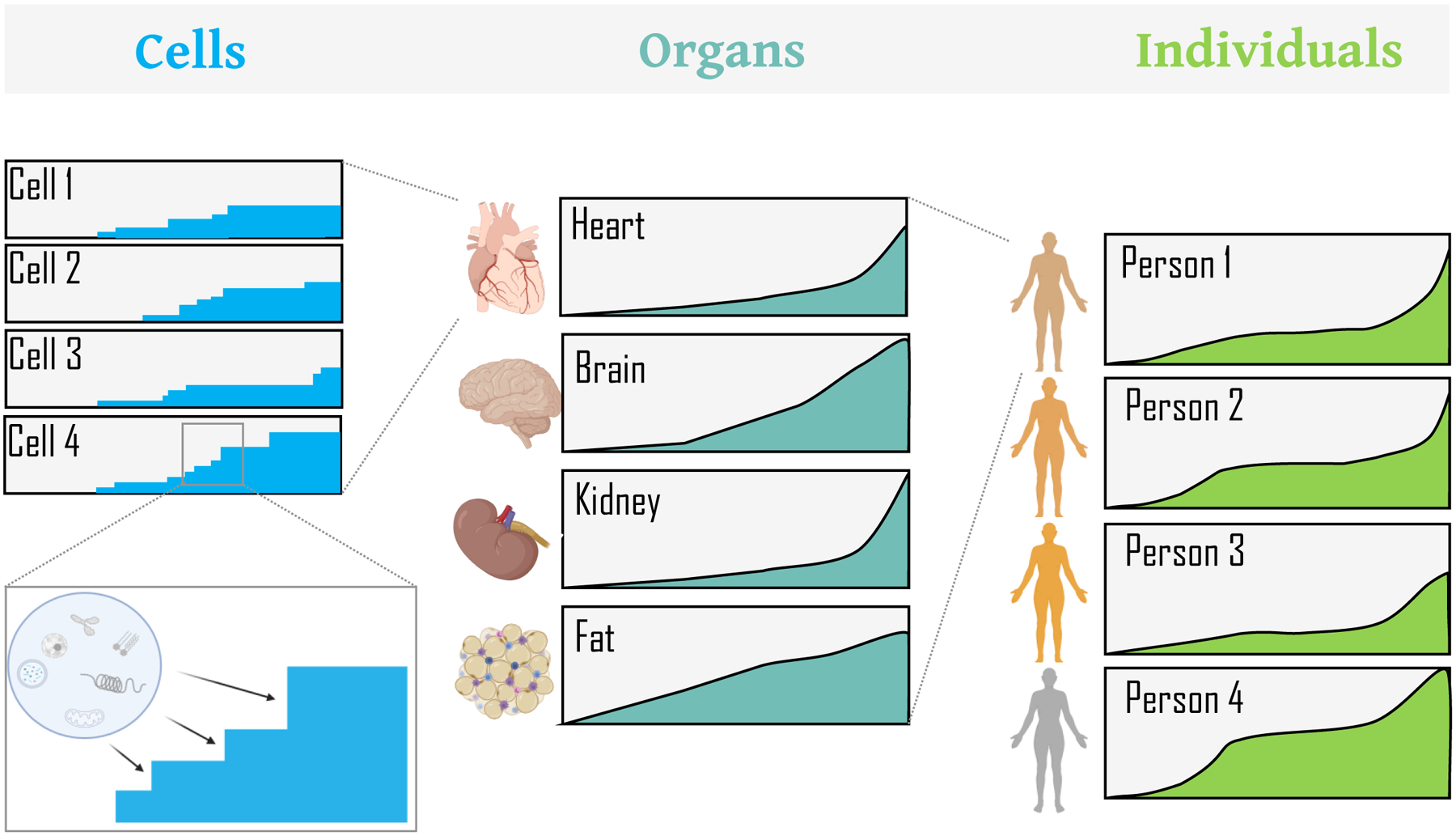
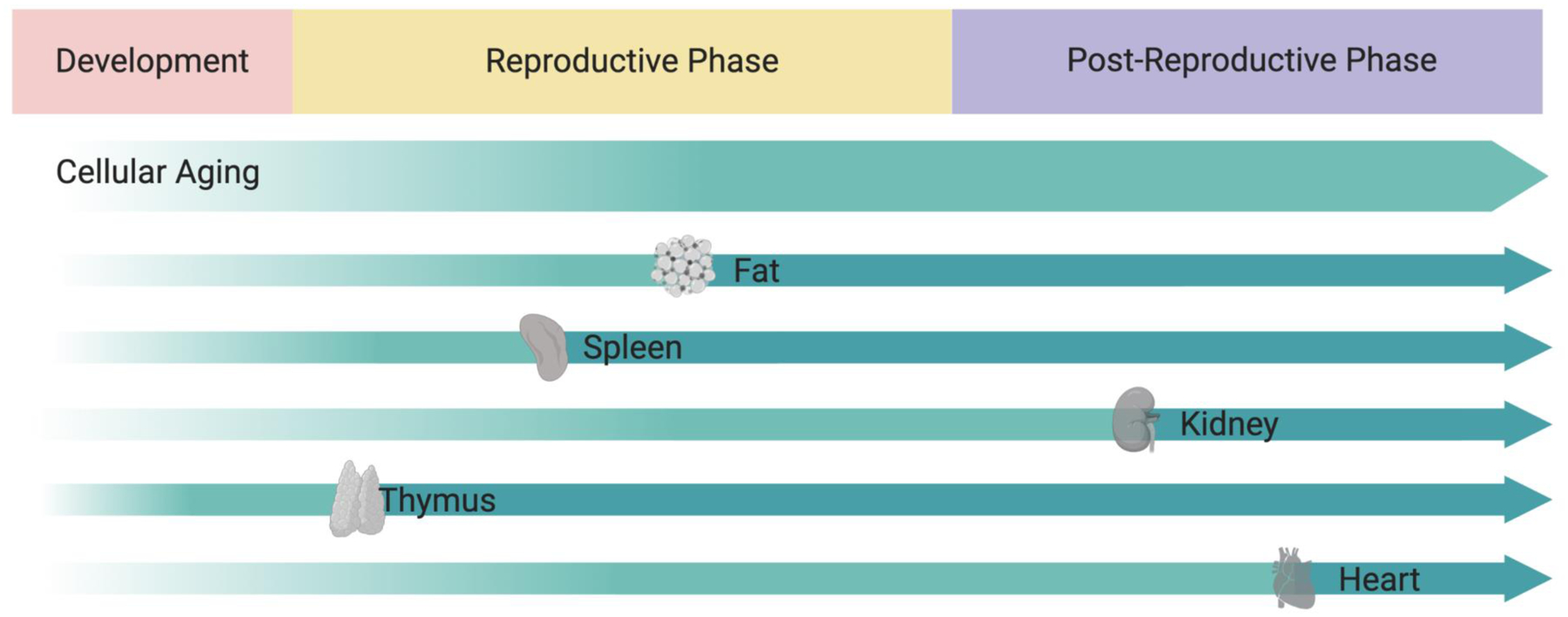
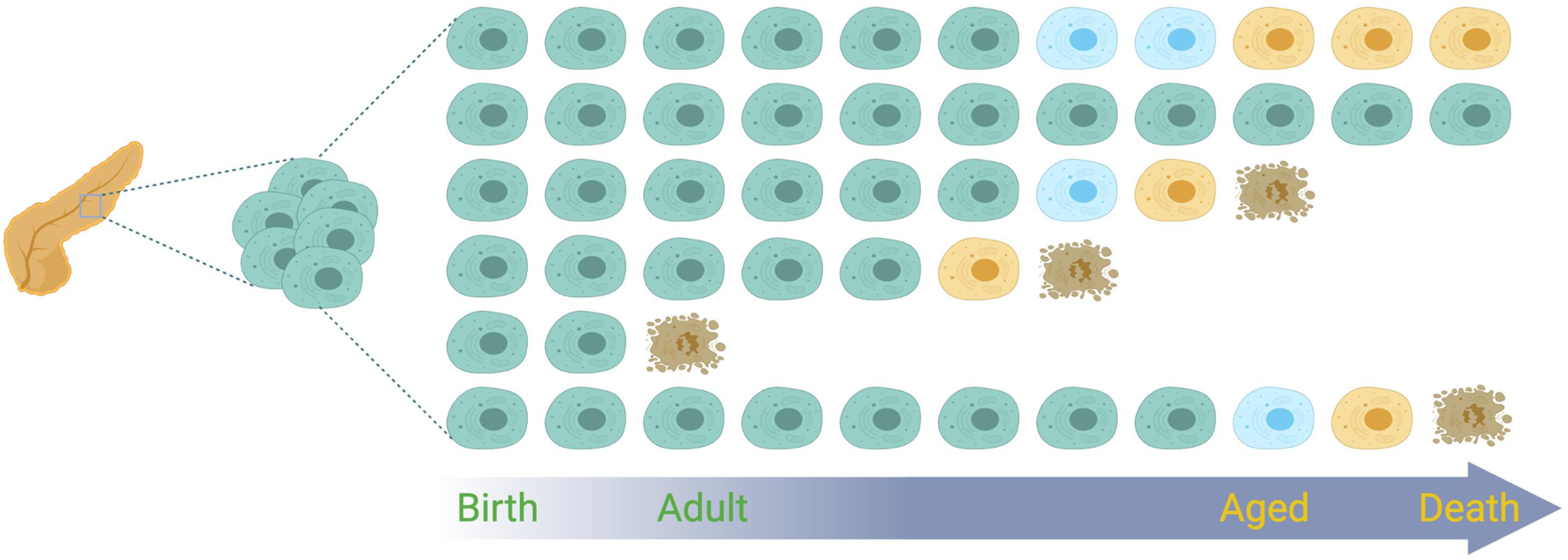
Aging has largely been defined by analog measures of organ and organismal dysfunction. This has led to the characterization of aging processes at the molecular and cellular levels that underlie these gradual changes. However, current knowledge does not fully explain the growing body of data emerging from large epidemiological, systems biology, and single cell studies of entire organisms pointing to varied rates of aging between individuals (different functionality and lifespan), across lifespan (asynchronous aging), and within an organism at the tissue and organ levels (aging mosaicism). Here we consider these inhomogeneities in the broader context of the rate of aging and from the perspective of underlying cellular changes. These changes reflect genetic, environmental, and stochastic factors that cells integrate to adopt new homeostatic, albeit less functional, states, such as cellular senescence. In this sense, cellular aging can be viewed, at least in part, as a quantal process we refer to as "digital aging". Nevertheless, analog declines of tissue dysfunction and organ failure with age could be the sum of underlying digital events. Importantly, cellular aging, digital or otherwise, is not uniform across time or space within the organism or between organisms of the same species. Certain tissues may exhibit earliest signs of cellular aging, acting as drivers for organismal aging as signals from those driver cells within those tissues may accelerate the aging of other cells locally or even systemically. Advanced methodologies at the systems level and at the single cell level are likely to continue to refine our understanding to the processes of how cells and tissues age and how the integration of those processes leads to the complexities of individual, organismal aging.
Figures
References
-
- Medawar PB (1952) An Unsolved Problem of Biology. H. K. Lewis, London.
-
- Williams GC (1957) Pleiotropy, natural selection, and the evolution of senescence. Evolution, 11: 398–411.
-
- Kirkwood TB (1977) Evolution of ageing. Nature, 270: 301–304. - PubMed
Publication types
MeSH terms
Grants and funding
LinkOut - more resources
Full Text Sources
Medical
