

The utility of computed tomography to evaluate thoracic complications after cardiopulmonary resuscitation
- PMID: 34223300
- PMCID: PMC8244247
- DOI: 10.1016/j.resplu.2020.100017
The utility of computed tomography to evaluate thoracic complications after cardiopulmonary resuscitation
Abstract
Background: Cardiopulmonary resuscitation (CPR) in adults following non-traumatic out of hospital cardiac arrest (OHCA) can cause thoracic complications including rib fractures, sternal fractures, and pneumothorax. Post-CPR complication rates are poorly studied and the optimum imaging modality to detect these complications post-resuscitation has not been established.
Methods: We performed a retrospective review of adult patients transported to a single, urban, academic hospital following atraumatic OHCA between September 2015 and January 2020. Patients who achieved sustained return of spontaneous circulation (ROSC) and who underwent computed tomography (CT) imaging of the chest following radiographic chest x-ray were included in the analyses. Patient demographics and prehospital data were collected. Descriptive statistics and multivariate logistic regression analysis were performed. Sensitivity and specificity of chest x-ray for the detection of thoracic injury in this population were estimated.
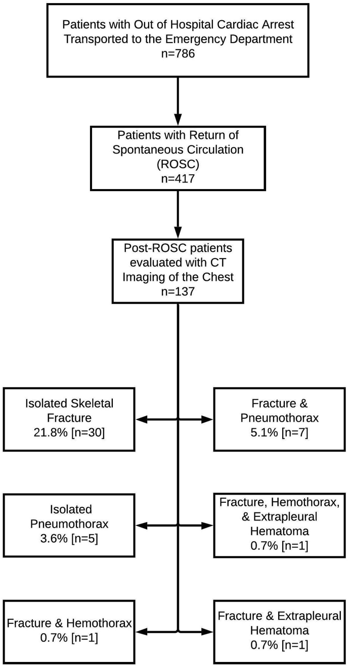
Results: 786 non-traumatic OHCA patients were transported to the ED, 417 of whom obtained sustained ROSC and were admitted to the hospital (53%). 137 (32.9%) admitted patients underwent CT imaging of the chest in the ED. Of these imaged patients median age was 62 years old (IQR 53-70) with 54.0% female and 38.0% of patients having received bystander CPR. 40/137 (29.2%) patients had skeletal fractures noted on CT imaging and 12/137 (8.8%) had pneumothorax present on CT imaging. X-ray yielded a sensitivity of 7.5% for rib fracture and 50% for pneumothorax with a specificity of 100% for both. Logistic regression analysis revealed no significant association between age, sex, bystander CPR, or resuscitation length with thoracic fractures or pneumothorax.
Conclusions: Complications from OHCA CPR were high with 29.2% of CT imaged patients having rib fractures and 8.8% having pneumothoraces. X-ray had poor sensitivity for these post-resuscitation complications. Post-CPR CT imaging of the chest should be considered for detecting post-CPR complications.
Keywords: Chest compression; Out of hospital CPR; Pneumothorax; Return of spontaneous circulation.
© 2020 The Author(s).
Conflict of interest statement
The authors have no conflicts of interest to disclose.
Figures
References
-
- Baringer J.R., Salzman E.W., Jones W.A. External cardiac massage. N Engl J Med. 1961;265(2):62–65. July 13. - PubMed
-
- Kouwenhoven W.B., Jude J.R., Knickerbocker G.G. CLOSED-CHEST cardiac massage. J Am Med Assoc. 1960;173(10):1064–1067. July 9. - PubMed
-
- Kleinman Monica E., Brennan Erin E., Goldberger Zachary D. Part 5: adult basic life support and cardiopulmonary resuscitation quality. Circulation. 2015;132(18_suppl_2):S414–S435. November 3. - PubMed
-
- Kleinman M.E., Goldberger Z.D., Rea T. American heart association focused update on adult basic life support and cardiopulmonary resuscitation quality: an update to the American heart association guidelines for cardiopulmonary resuscitation and emergency cardiovascular care. Circulation. 2017;(1):137. 2018;January 2. - PubMed
-
- Tsitlik J.E., Weisfeldt M.L., Chandra N. Elastic properties of the human chest during cardiopulmonary resuscitation. Crit Care Med. 1983;11(9):685–692. September. - PubMed
LinkOut - more resources
Full Text Sources
