Review
. 2021 Aug 10;65(3):467-480.
doi: 10.1042/EBC20200128.

3D cell aggregate printing technology and its applications

Affiliations
Review

3D cell aggregate printing technology and its applications

Seunggyu Jeon et al. Essays Biochem. .

Abstract

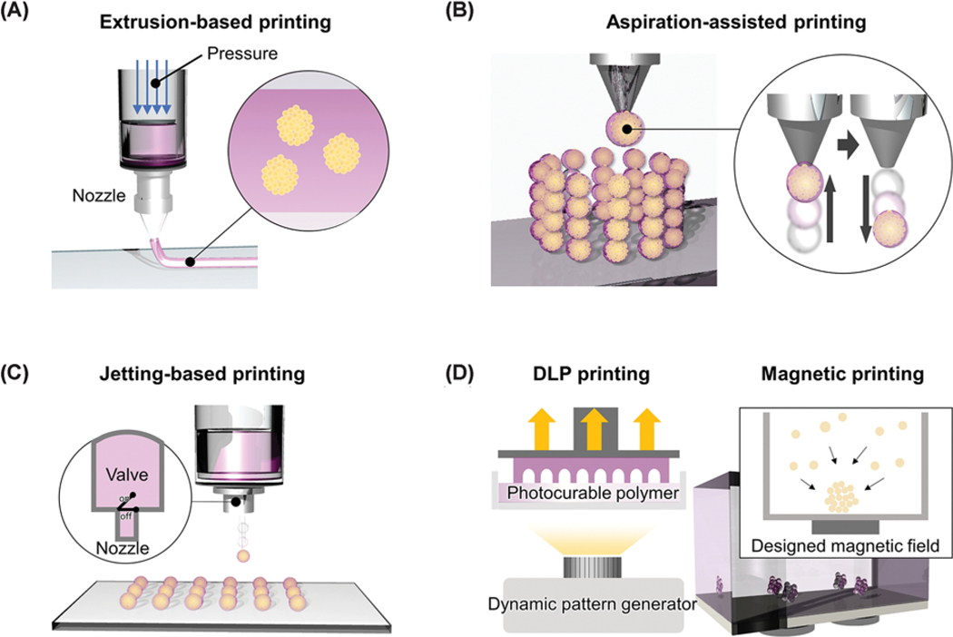
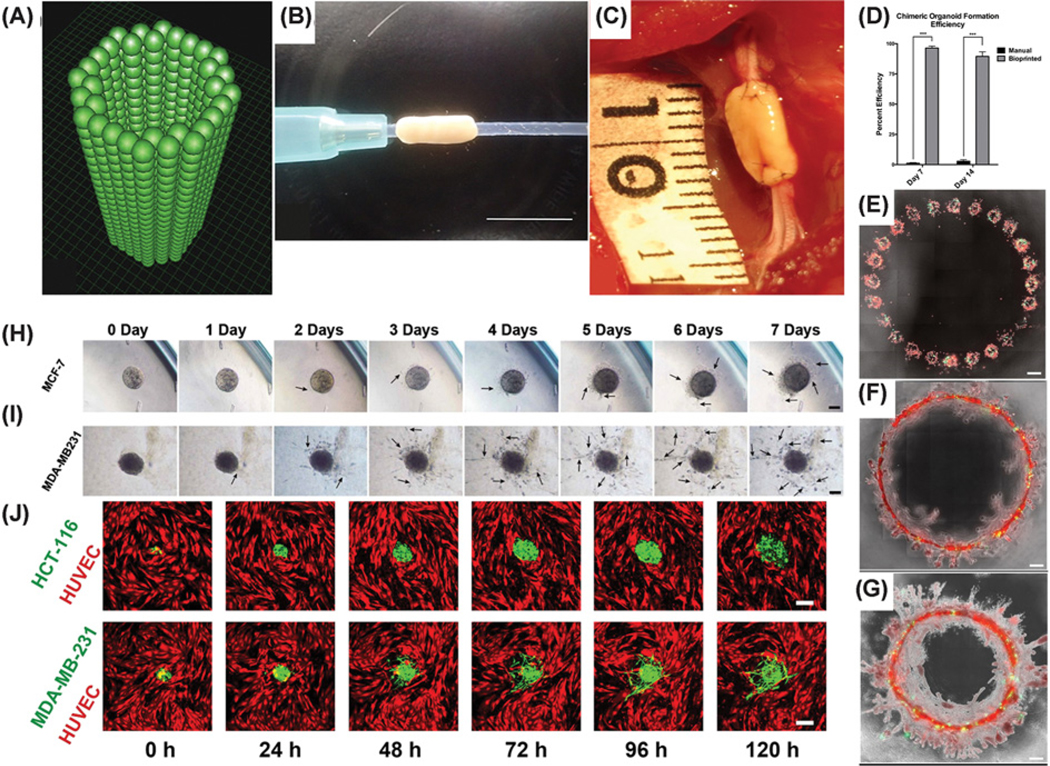
Various cell aggregate culture technologies have been developed and actively applied to tissue engineering and organ-on-a-chip. However, the conventional culture technologies are labor-intensive, and their outcomes are highly user dependent. In addition, the technologies cannot be used to produce three-dimensional (3D) complex tissues. In this regard, 3D cell aggregate printing technology has attracted increased attention from many researchers owing to its 3D processability. The technology allows the fabrication of 3D freeform constructs using multiple types of cell aggregates in an automated manner. Technological advancement has resulted in the development of a printing technology with a high resolution of approximately 20 μm in 3D space. A high-speed printing technology that can print a cell aggregate in milliseconds has also been introduced. The developed aggregate printing technologies are being actively applied to produce various types of engineered tissues. Although various types of high-performance printing technologies have been developed, there are still some technical obstacles in the fabrication of engineered tissues that mimic the structure and function of native tissues. This review highlights the central importance and current technical level of 3D cell aggregate printing technology, and their applications to tissue/disease models, artificial tissues, and drug-screening platforms. The paper also discusses the remaining hurdles and future directions of the printing processes.

Keywords: 3D bioprinting; cell aggregate; tissue engineering; tissue/disease model.


Figures

Figure 1.
Figure 1.. Schematic illustrations of 3D cell aggregate printing technologies
Extrusion-based printing (A), aspiration-assisted printing (B), ink-jet-based printing (C), other printing technologies (DLP and magnetic printing) (D). Abbreviation: DLP, digital light processing.
Figure 2.
Figure 2.. 3D extrusion-based and aspiration-assisted cell aggregate printing technologies
(A) Illustration of direct cell aggregate printing process using an extrusion-based printer. (B) Photograph of the construct printed by deposition of agarose cylinders and cell aggregates [20]. (C) Schematic showing the cell printing and induction of cell aggregates at the printed sites. (D) Induced cell aggregates in a dextran (DEX) solution using a two-phase system with polyethylene glycol (PEG) (scale bar: 200 μm) [24]. (E) Schematic to illustrate the aspiration-assisted printing process of cell aggregates into microneedles. (F) Design (left) and printed result (right) of a heterogeneous tubular structure consisting of two types of cell aggregates [27]. (G) Step-by-step photographs of 3D positioning of cell aggregates into a hydrogel [14]. (B,D,F,G) were reproduced with permission from [14,20,24,27].
Figure 3.
Figure 3.. Other 3D aggregate printing technologies
(A) Step-by-step illustration of the hanging drop culture method by jetting cell-laden droplets on to the lid of a Petri dish [32]. (B) Schematic illustration of the singularization of a cell aggregate using an imaging and vacuum shutter system [34]. (C) Photograph of a 3D scaffold with individually positioned cell aggregates [35]. (D) Schematic drawings showing the process of forming a concave pocket using a 3D printed mold (i,ii,iii) and the role of a small inlet for cell seeding and aggregate culture (iv,v) [44]. (E) Schematic and results of controlling cell composition within a single cell aggregate by applying FACS (scale bar: 200 μm) [49]. The figures were reproduced with permission from [32,34,35,44,49]. Abbreviation: FACS, fluorescence-activated cell sorting.
Figure 4.
Figure 4.. 3D cell aggregates fabricated for blood vessel reconstruction
Blood vessel tubular model: (A) a scaffold-free vascular graft is generated from MCSs, (B) Masson’s Trichrome staining image of the vascular graft in a short axis cross-section, (C) Cell Tracker Red-labeled image of the lumen area after implantation [26]. Cardiac patch model: (D) transplanted cardiac patch on to the rat heart, (E) Hematoxylin and Eosin stain (H&E, white arrows: erythrocytes, scale bar: 400 μm), (F) human nuclear antigen (HNA, magenta), wheat germ agglutinin (WGA, green), and DAPI (blue, white arrows: human cells in native rat myocardium, white dotted line: cardiac patch (left) from native rat myocardium (right), scale bar: 40 μm) [50]. Cardiac tube model: (G) fabricated tubular cardiac constructs, (H) terminal deoxynucleotidyl transferase dUTP nick-end labeling (TUNEL) image of a cell aggregate (×20, scale bar: 100 μm), (I) TUNEL image of the tubular construct (×4, scale bar: 100 μm) [51]. The figures were reproduced with permission from [26,50,51].
Figure 5.
Figure 5.. 3D cell aggregates fabricated to induce nerve conduit and cancer model
Conduit model: (A) pre-designed 3D tube-like structure. (B) Fabricated 3D conduit (scale bar: 10 mm). (C) Image of the conduit grafted into the rat nerve [16]. Breast cancer cell aggregates: (D) chimeric cell aggregate formation efficiency (***P<0.001). Printed circular pattern (MCF-12A (red) and MDA-MB-468 (green) cells): (E) day 3. (F) day 7. (G) day 21 (scale bars: 500 μm) [58]. Time-lapse images for different cell spheroid invasions inside a collagen gel: (H) MCF-7. (I) MDA-MB-231 (scale bars: 100 μm). (J) Time-lapse confocal images for different cell spheroids of transendothelial migration inside a collagen gel (HCT-116 (green), MDA-MB-231 (green), and HUVEC (red)) [59]. The figures were reproduced with permission from [16,58,59].
Figure 6.
Figure 6.. 3D cell aggregates fabricated to reconstruct trachea, tendon, and liver tissues
Trachea-like model: (A) transplantation image. (B) Histological and immunohistological staining with anti-CD31 antibodies (arrows: capillary-like tube formation, scale bar: 200 μm). (C) Histological and immunohistological staining with anti-pan-cytokeratin antibodies (arrows: trachea epithelial layer, scale bars: 400 μm) [17]. Tendon-like model: (D) images showing changes in construct shape with prolonged culture (scale bars: 2 mm). (E) 4 weeks. (F) 8 weeks. (G) Tenascin C staining image of the tension-loaded group (scale bar: 20 μm). (H) Scleraxis staining image of the tension-loaded group (scale bar: 10 μm) [61]. Liver-like model: (I) H&E-stained cross-sections at day 7 of a 3D printed liver-like construct. (J) Fluorescence image of the liver-like tissue on day 7 (hHep: green, HUVEC: red, scale bars: 200 μm). (K) Transplanted liver-like tissue graft. (L) Immunostaining image of human albumin (scale bar: 200 μm). (M) Immunostaining image of human CYP3A4 (scale bar: 200 μm) [63]. The figures were reproduced with permission from [17,61].

Similar articles

Cited by

References

    1. Laschke MW and Menger MD (2017) Life is 3D: boosting spheroid function for tissue engineering. Trends Biotechnol. 35, 133–144, 10.1016/j.tibtech.2016.08.004 - DOI - PubMed
    1. Wörsdörfer P, Dalda N, Kern A, Krüger S, Wagner N, Kwok CK et al. (2019) Generation of complex human organoid models including vascular¨ networks by incorporation of mesodermal progenitor cells. Sci. Rep. 9, 15663, 10.1038/s41598-019-52204-7 - DOI - PMC - PubMed
    1. Mattix B, Olsen TR, Gu Y, Casco M, Herbst A, Simionescu DT et al. (2014) Biological magnetic cellular spheroids as building blocks for tissue engineering. Acta Biomater. 10, 623–629, 10.1016/j.actbio.2013.10.021 - DOI - PMC - PubMed
    1. Shoemaker JT, Zhang W, Atlas SI, Bryan RA, Inman SW and Vukasinovic J. (2020) A 3D cell culture organ-on-a-chip platform with a breathable hemoglobin analogue augments and extends primary human hepatocyte functions in vitro. Front. Mol. Biosci. 7, 296, 10.3389/fmolb.2020.568777 - DOI - PMC - PubMed
    1. Lin B, Miao Y, Wang J, Fan Z, Du L, Su Y. et al. (2016) Surface Tension Guided Hanging-Drop: producing controllable 3D spheroid of high-passaged human dermal papilla cells and forming inductive microtissues for hair-follicle regeneration. ACS Appl. Mater. Interfaces 8, 5906–5916, 10.1021/acsami.6b00202 - DOI - PubMed

Publication types

MeSH terms

LinkOut - more resources