Review
. 2022 Feb;18(2):360-376.
doi: 10.1002/alz.12389. Epub 2021 Jul 5.

Diversity of transcriptomic microglial phenotypes in aging and Alzheimer's disease

Affiliations
Review

Diversity of transcriptomic microglial phenotypes in aging and Alzheimer's disease

Delphine Boche et al. Alzheimers Dement. 2022 Feb.

Abstract

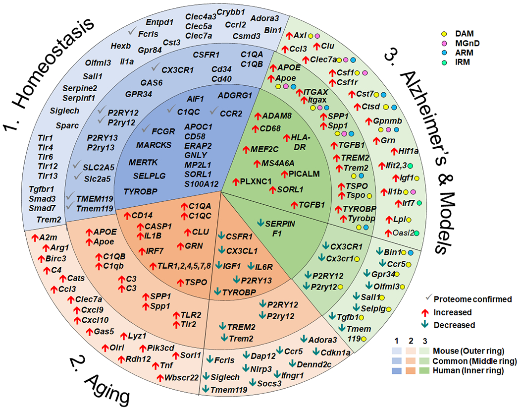
The morphological plasticity of microglia has fascinated neuroscientists for 100 years. Attempts to classify functional phenotypes are hampered by similarities between endogenous brain microglia and peripheral myeloid cells that can enter the brain under pathological conditions. Recent advances in single-cell -omic methodologies have led to an explosion of data regarding gene expression in microglia. Herein, we review the diversity of microglial phenotypes in healthy brains, aging, and Alzheimer's disease (AD); identify knowledge gaps in the body of evidence; and suggest areas in which new knowledge would be useful. Data from human samples and mouse models are compared and contrasted. Understanding the molecular complexity of the microglial response repertoire will suggest new avenues for therapeutic treatments in AD.

Keywords: gene expression; immunity; microarray; microglia; neurodegeneration; sequencing.


Conflict of interest statement

Declarations of interest: none

Figures

Figure 1.
Figure 1.
Summary of select gene expression changes in health, aging and Alzheimer’s disease. The circle represents gene expression. Data from mice are presented in the outer ring (light shading), while data from human are presented in the interior circle (darkest shading). Gene changes that are expressed by both human and mouse are presented in the middle ring with medium shading. Genes associated with homeostatic microglia are presented within the blue wedge (1) with the gene expression changes confirmed at the proteomic level indicated with a grey check mark. Changes in gene expression accompanying aging are presented in the beige wedge (2). Genes that are increased with aging relative to young adult are indicated with red up arrows, while genes that decrease are indicated with green down arrows. Changes in gene expression accompanying Alzheimer’s disease or Alzheimer-like pathology in mouse models are presented in the green wedge (3). In humans, increases and decreases compared with participants with no cognitive impairment are displayed in the green inner ring with red up or green down arrows, respectively. Within the wedge and ring for mouse models with Alzheimer-like pathology, the genes associated with the activated microglial subtypes DAM, MGnD, ARM or IRM are identified with color dots and the increase or decrease expression relative to the homeostatic profile indicated with red or green arrows. Not all gene changes can be depicted in the diagram.

References

    1. Ginhoux F, Greter M, Leboeuf M, Nandi S, See P, Gokhan S, Mehler MF, Conway SJ, Ng LG, Stanley ER, Samokhvalov IM, Merad M. Fate mapping analysis reveals that adult microglia derive from primitive macrophages. Science 2010; 330: 841–5. - PMC - PubMed
    1. Kierdorf K, Erny D, Goldmann T, Sander V, Schulz C, Perdiguero EG, Wieghofer P, Heinrich A, Riemke P, Holscher C, Muller DN, Luckow B, Brocker T, Debowski K, Fritz G, Opdenakker G, Diefenbach A, Biber K, Heikenwalder M, Geissmann F, Rosenbauer F, Prinz M. Microglia emerge from erythromyeloid precursors via Pu.1- and Irf8-dependent pathways. Nat Neurosci 2013; 16: 273–80. - PubMed
    1. McCarter JC. A silver carbonate staining method for oligodendrocytes and microglia for routine use. Am J Pathol. 1940. Mar;16(2):233–235. - PMC - PubMed
    1. Mori S, Leblond CP. Identification of microglia in light and electron microscopy. J Comp Neurol 1969; 135: 57–80. - PubMed
    1. Grassivaro F, Menon R, Acquaviva M, Ottoboni L, Ruffini F, Bergamaschi A, Muzio L, Farina C, Martino G. Convergence between microglia and peripheral macrophages phenotype during development and neuroinflammation. J Neurosci 2020; 40: 784–95. - PMC - PubMed

Publication types