Multicenter Study
. 2021 Dec;42(6):664-673.
doi: 10.1080/13816810.2021.1946704. Epub 2021 Jul 5.

Expanding the clinical phenotype in patients with disease causing variants associated with atypical Usher syndrome

Affiliations
Multicenter Study

Expanding the clinical phenotype in patients with disease causing variants associated with atypical Usher syndrome

Austin D Igelman et al. Ophthalmic Genet. 2021 Dec.

Abstract

Atypical Usher syndrome (USH) is poorly defined with a broad clinical spectrum. Here, we characterize the clinical phenotype of disease caused by variants in CEP78, CEP250, ARSG, and ABHD12.Chart review evaluating demographic, clinical, imaging, and genetic findings of 19 patients from 18 families with a clinical diagnosis of retinal disease and confirmed disease-causing variants in CEP78, CEP250, ARSG, or ABHD12.CEP78-related disease included sensorineural hearing loss (SNHL) in 6/7 patients and demonstrated a broad phenotypic spectrum including: vascular attenuation, pallor of the optic disc, intraretinal pigment, retinal pigment epithelium mottling, areas of mid-peripheral hypo-autofluorescence, outer retinal atrophy, mild pigmentary changes in the macula, foveal hypo-autofluorescence, and granularity of the ellipsoid zone. Nonsense and frameshift variants in CEP250 showed mild retinal disease with progressive, non-congenital SNHL. ARSG variants resulted in a characteristic pericentral pattern of hypo-autofluorescence with one patient reporting non-congenital SNHL. ABHD12-related disease showed rod-cone dystrophy with macular involvement, early and severe decreased best corrected visual acuity, and non-congenital SNHL ranging from unreported to severe.This study serves to expand the clinical phenotypes of atypical USH. Given the variable findings, atypical USH should be considered in patients with peripheral and macular retinal disease even without the typical RP phenotype especially when SNHL is noted. Additionally, genetic screening may be useful in patients who have clinical symptoms and retinal findings even in the absence of known SNHL given the variability of atypical USH.

Keywords: ABHD12; ARSG; Atypical usher syndrome; CEP78; cep250.


Conflict of interest statement

Disclosure of interest: The authors report no conflict of interest.

Figures

Figure 1:
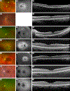
Figure 1:. Representative multimodal imaging of patients with CEP78-related disease
CEP78–18807 (A – C), CEP78–2 (D, E), CEP78–3 (F – H), CEP78–4 (I – K), CEP78–5 (L – N), CEP78–87042 (O – Q) CEP78–7 (R – T). CFP (A, D, F, I, L, O, R), FAF (B, G, J, M, P, S), OCT sections (C, E, H, K, N, Q, T) demonstrating the spectrum of disease. Subtype 1 depicts an RP like phenotype including vascular attenuation, pallor of the optic disc, and RPE mottling on CFP (A, D, F, I, L), areas of mid peripheral hypo- autofluorescence ranging from mild to severe on FAF (B, G, J, M), and outer retinal atrophy on OCT (C, E, H, K). Common findings in subtype 1 include foveal hypo-autofluorescence (P, S) and granularity of the ellipsoid zone (Q, T). Abbreviations: CFP, color fundus photos; FAF, fundus auto-fluorescence; OCT, optical coherence tomography; ERM, epiretinal membrane.
Figure 2:
Figure 2:. Representative multimodal imaging of patients with CEP250-related disease
CEP250–1 (A – C), CEP250–2 (D – F), CEP250–3 (G – I). CFP (A, D, G), FAF (B, E, H), OCT sections (C, F, I) demonstrating the spectrum of disease. CFP revealed normal findings in all patients (A, D, G). FAF ranged from normal (E) to areas of subtle hyper-autofluorescence in the periphery (B) and in the peripapillary region (H). OCT findings showed outer retinal atrophy in all three patients including thinning of the outer nuclear layer (C) and subtle disruption of the ellipsoid zone and interdigitation zone (I). Abbreviations: CFP, color fundus photos; FAF, fundus auto-fluorescence; OCT, optical coherence tomography.
Figure 3:
Figure 3:. Representative multimodal imaging of patients with ARSG-related disease
ARSG-1 (A – C), ARSG-2 (D – F). ARSG-29692 (G – I). CFP (A, D, G), FAF (B, E, H), OCT sections (C, F, I) demonstrating the spectrum of disease. CFP showed parafoveal and mid-peripheral RPE atrophy, intraretinal pigment, and optic disc pallor (A, D, G). FAF of all patients demonstrated pericentral and mid-peripheral hypo-autofluorescence (B, E, H). ARSG-29692 revealed advanced disease and macular involvement with central hypo-autofluorescence (H), ARSG-2 showed a parafoveal ring of hypo-autofluorescence (E), and ARSG-1 demonstrated a parafoveal hyper-autofluorescent ring (B). OCT revealed extensive outer retinal atrophy involving the foveal in ARSG-29692 (I), and foveal sparing atrophy in ARSG-1 and ARSG-2 (C, F). Abbreviations: CFP, color fundus photos; FAF, fundus auto-fluorescence; OCT, optical coherence tomography.
Figure 4:
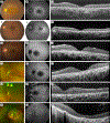
Figure 4:. Representative multimodal imaging of patients with ABHD12-related disease
ABHD12–1 (A – C), ABHD12–2 (D – F), ABHD12–3 (G – I), ABHD12–4 (J – L), ABHD12–5 (M – O), ABHD12–6 (P – RCFP (A, D, G, J, M, P), FAF (B, E, H, K, N, Q), OCT sections (C, F, I, L, O, R) demonstrating the spectrum of disease. CFP showed macular changes in all patients ranging from mild granular changes (D) to significant RPE atrophy (P). FAF revealed macular involvement ranging from central hypo-autofluorescence (H, K) to severe global hypo-autofluorescence (Q). OCT showed fovea-involving outer retinal atrophy and as sub-retinal deposits. Abbreviations: CFP, color fundus photos; FAF, fundus auto-fluorescence; OCT, optical coherence tomography.

References

    1. Mathur P, Yang J. Usher syndrome: Hearing loss, retinal degeneration and associated abnormalities. Biochim Biophys Acta. 2015;1852(3):406–20. - PMC - PubMed
    1. Nolen RM, Hufnagel RB, Friedman TB, Turriff AE, Brewer CC, Zalewski CK, et al. Atypical and ultra-rare Usher syndrome: a patients with Usher syndrome type 1. J Hum Genet. 2010;55(12):796–800. - PubMed
    1. Bashir R, Fatima A, Naz S. A frameshift mutation in SANS results in atypical Usher syndrome. Clin Genet. 2010;78(6):601–3. - PMC - PubMed
    1. Fu Q, Xu M, Chen X, Sheng X, Yuan Z, Liu Y, et al. CEP78 is mutated in a distinct type of Usher syndrome. J Med Genet. 2017;54(3):190–5. - PMC - PubMed
    1. Liu X-Z, Hope C, Walsh J, Newton V, Ke XM, Liang CY, et al. Mutations in the myosin VIIA gene cause a wide phenotypic spectrum, including atypical Usher syndrome. The American Journal of Human Genetics. 1998;63(3):909–12. - PMC - PubMed

Publication types

MeSH terms