. 2021 Jul 26;67(3):83-94.
doi: 10.1530/JME-21-0030.

The bromodomain inhibitor JQ1+ reduces calcium-sensing receptor activity in pituitary cell lines

Affiliations

The bromodomain inhibitor JQ1+ reduces calcium-sensing receptor activity in pituitary cell lines

Kate E Lines et al. J Mol Endocrinol. .

Abstract

Corticotrophinomas represent 10% of all surgically removed pituitary adenomas, however, current treatment options are often not effective, and there is a need for improved pharmacological treatments. Recently, JQ1+, a bromodomain inhibitor that promotes gene transcription by binding acetylated histone residues and recruiting transcriptional machinery, has been shown to reduce proliferation in a murine corticotroph cell line, AtT20. RNA-Seq analysis of AtT20 cells following treatment with JQ1+ identified the calcium-sensing receptor (CaSR) gene as significantly downregulated, which was subsequently confirmed using real-time PCR and Western blot analysis. CaSR is a G protein-coupled receptor that plays a central role in calcium homeostasis but can elicit non-calcitropic effects in multiple tissues, including the anterior pituitary where it helps regulate hormone secretion. However, in AtT20 cells, CaSR activates a tumour-specific cAMP pathway that promotes ACTH and PTHrP hypersecretion. We hypothesised that the Casr promoter may harbour binding sites for BET proteins, and using chromatin immunoprecipitation (ChIP)-sequencing demonstrated that the BET protein Brd3 binds to the promoter of the Casr gene. Assessment of CaSR signalling showed that JQ1+ significantly reduced Ca2+e-mediated increases in intracellular calcium (Ca2+i) mobilisation and cAMP signalling. However, the CaSR-negative allosteric modulator, NPS-2143, was unable to reduce AtT20 cell proliferation, indicating that reducing CaSR expression rather than activity is likely required to reduce pituitary cell proliferation. Thus, these studies demonstrate that reducing CaSR expression may be a viable option in the treatment of pituitary tumours. Moreover, current strategies to reduce CaSR activity, rather than protein expression for cancer treatments, may be ineffective.

Keywords: G protein-coupled receptor; corticotrophinoma; epigenetic modification; pituitary tumourigenesis.


Figures
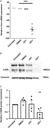
Figure 1 Calcium-sensing receptor
(CaSR) is downregulated by JQ1+ treatment. (A) qRT-PCR analysis of CaSR following exposure to cell culture media, DMSO, JQ1− and JQ1+ for 96 h in AtT20 cells. (B) Representative Western blot of CaSR in AtT20 cells following exposure to DMSO, JQ1− and JQ1+ for 96 h. (C) Densitometry analysis of protein expression following treatment with DMSO, JQ1− and JQ1+ for 96 h. CaSR protein expression was expressed relative to calnexin. Data are expressed as mean ± s.e.m. in panels A and C. Statistical analyses compared to DMSO-treated cells in all panels: ***P < 0.001, **P < 0.01.
Figure 2
Figure 2
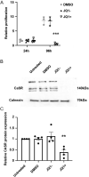
JQ1+ does not reduce proliferation of AtT20 cells after 24 h but does decrease calcium-sensing receptor (CaSR) protein expression. (A) Relative proliferation of AtT20 cells following exposure to DMSO, JQ1− and JQ1+ for 24 and 96 h. Data were expressed relative to cell numbers at day 0. (B) Representative Western blot of CaSR in AtT20 cells following exposure to DMSO, JQ1− and JQ1+ for 24 h. (C) Densitometry analysis of protein expression following treatment with DMSO, JQ1− and JQ1+ for 24 h. CaSR protein expression was expressed relative to calnexin. Data are expressed as mean ± s.e.m. in panels A and C. Statistical analyses compared to DMSO treatment in panel A, ***P < 0.001, and statistical analysis compared to DMSO-treated cells in panel C, **P < 0.005.
Figure 3
Figure 3
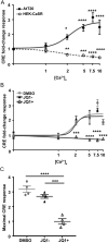
JQ1+ reduces cAMP signalling in AtT20 cell lines. (A) Ca2+e-induced cAMP-response element (CRE) luciferase reporter responses in AtT20 and HEK-CaSR cell lines. Elevations in Ca2+e concentrations increase CRE luciferase in AtT20 cells and reduce luciferase in HEK-CaSR cells. (B) Ca2+e-induced CRE luciferase reporter responses in AtT20 cell lines following exposure to DMSO, JQ1− and JQ1+ for 24 h. (C) Maximal CRE luciferase responses from panel B. Data are expressed as mean ± s.e.m. in all panels. Statistical analyses compared to basal responses in each cell line in panel A and to DMSO-treated cells in panel B. Firefly values ranged from 6027 to 981,659 luminescent units and renilla from 1027 to 5031 luminescent units. ****P < 0.0001, ***P < 0.001, **P < 0.01, *P < 0.05.
Figure 4
Figure 4
JQ1+ reduces calcium-sensing receptor (CaSR)-mediated calcium mobilisation in AtT20 cell lines. (A) Ca2+ e-induced Fluo-4 intracellular calcium mobilisation assays in AtT20 cells following exposure to DMSO, JQ1− and JQ1+ for 24 h. (B) Area under the curve (AUC) of data in A. (C) Maximal Ca2+i responses and (D) EC50 values obtained in Fluo-4 intracellular calcium mobilisation assays shown in panel A. Data are expressed as mean ± s.e.m. in panels A and B, and mean ± 95% CIs in panel C. Statistical analyses compared between groups in panel B, ***P < 0.001, *P < 0.05.
Figure 5
Figure 5
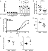
NPS-2143 has no effect on AtT20 proliferation but does reduce calcium-sensing receptor (CaSR)-mediated cAMP and Ca2+i signalling in AtT20 cells. (A) Effect of NPS-2143 (0–1000 nM) on proliferation of AtT20 cells 2 days and 7 days post-treatment. Data were expressed relative to proliferation at day 1. Statistical analyses comparing proliferation between day 2 and day 7 in each group. There was no significant difference between cell proliferation in cells exposed to different concentrations of NPS-2143. Raw fluorescence units ranged between 17,096 and 69,941 for day 2 and 19,935 and 222,377 for day 7. (B) cAMP-response element (CRE) luciferase reporter responses at 5 mM Ca2+e in AtT20 cells exposed to DMSO or 20 nM NPS-2143 for 12 h. Data were expressed relative to responses in DMSO-treated cells. (C) Ca2+e-induced Fluo-4 intracellular calcium mobilisation assays in AtT20 cells following exposure to DMSO or 20 nM NPS-2143. Raw fluorescence units ranged between 1896 and 58,337. (D) Area under the curve (AUC) of data in C, (E) Maximal Ca2+i responses, and (F) EC50 values showing mean ± 95% CIs obtained in Fluo-4 intracellular calcium mobilisation assays shown in panel C. ****P < 0.0001, ***P < 0.001, **P < 0.01, *P < 0.05.

Similar articles

Cited by

References

    1. Asadi F, Farraj M, Sharifi R, Malakouti S, Antar S, Kukreja S. 1996. Enhanced expression of parathyroid hormone-related protein in prostate cancer as compared with benign prostatic hyperplasia. Human Pathology 27 1319–1323. (10.1016/s0046-8177(9690344-5) - DOI - PubMed
    1. Bibonne A, Néant I, Batut J, Leclerc C, Moreau M, Gilbert T. 2013. Three calcium-sensitive genes, fus, brd3 and wdr5, are highly expressed in neural and renal territories during amphibian development. Biochimica et Biophysica Acta 1833 1665–1671. (10.1016/j.bbamcr.2012.12.015) - DOI - PubMed
    1. Birkenkamp-Demtroder K, Hahn SA, Mansilla F, Thorsen K, Maghnouj A, Christensen R, Oster B, Orntoft TF. 2013. Keratin23 (KRT23) knockdown decreases proliferation and affects the DNA damage response of colon cancer cells. PLoS ONE 8 e73593. (10.1371/journal.pone.0073593) - DOI - PMC - PubMed
    1. Brown EM, MacLeod RJ. 2001. Extracellular calcium sensing and extracellular calcium signaling. Physiological Reviews 81 239–297. (10.1152/physrev.2001.81.1.239) - DOI - PubMed
    1. Cuevas-Ramos D, Lim DST, Fleseriu M. 2016. Update on medical treatment for Cushing’s disease. Clinical Diabetes and Endocrinology 2 16. (10.1186/s40842-016-0033-9) - DOI - PMC - PubMed

Publication types