. 2021 Aug 19;42(8):1079-1088.
doi: 10.1093/carcin/bgab059.

Appearance of peanut agglutinin in the blood circulation after peanut ingestion promotes endothelial secretion of metastasis-promoting cytokines

Affiliations

Appearance of peanut agglutinin in the blood circulation after peanut ingestion promotes endothelial secretion of metastasis-promoting cytokines

Weikun Wang et al. Carcinogenesis. .

Abstract

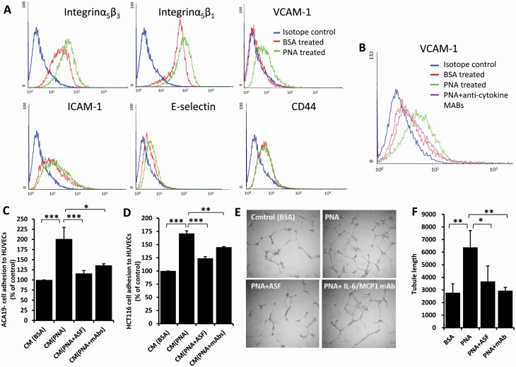
Peanut agglutinin (PNA) is a carbohydrate-binding protein in peanuts that accounts for ~0.15% peanut weight. PNA is highly resistant to cooking and digestion and is rapidly detectable in the blood after peanut consumption. Our previous studies have shown that circulating PNA mimics the actions of endogenous galactoside-binding protein galectin-3 by interaction with tumour cell-associated MUC1 and promotes circulating tumour cell metastatic spreading. The present study shows that circulating PNA interacts with micro- as well as macro-vascular endothelial cells and induces endothelial secretion of cytokines MCP-1 (CCL2) and IL-6 in vitro and in vivo. The increased secretion of these cytokines autocrinely/paracrinely enhances the expression of endothelial cell surface adhesion molecules including integrins, VCAM and selectin, leading to increased tumour cell-endothelial adhesion and endothelial tubule formation. Binding of PNA to endothelial surface MCAM (CD146), via N-linked glycans, and subsequent activation of PI3K-AKT-PREAS40 signalling is here shown responsible for PNA-induced secretion of MCP-1 and IL-6 by vascular endothelium. Thus, in addition to its influence on promoting tumour cell spreading by interaction with tumour cell-associated MUC1, circulating PNA might also influence metastasis by enhancing the secretion of metastasis-promoting MCP-1 and IL-6 from the vascular endothelium.


Figures

Figure 1.
Figure 1.
PNA induces MCP-1 and IL-6 secretion from micro- and macro-vascular endothelial cells in vitro and in mice. (A) HMVEC-Ls were treated with 4 μg/ml PNA or BSA for 24 h. The levels of cytokines in the culture media were analysed by cytokine profile array. PNA induces dose- (B, C and F, G) and time- (D, E and H, I) dependent increase of MCP-1 (B, D and F, H) and IL-6 (C, E and G, I) secretion from micro-vascular HMVEC-Ls (AE) and macro-vascular HUVECs (FI). (J and K) Nine mice were injected intravenously with 10 μg PNA and the blood from three mice at 0, 24 and 48 h were obtained and serum concentrations of MCP-1 and IL-6 in the blood were analysed. Data are presented as Mean ± SD of three independent experiments, each in triplicate. *p < 0.05, **p < 0.01, ***p < 0.001.
Figure 2.
Figure 2.
PNA-induced cytokine secretion increases cancer cell-endothelial adhesion and endothelial tubule formation. (A and B) PNA-induced cytokine secretion increases the expression of endothelial cell surface adhesion molecules. HUVECs were treated with 4 μg/ml PNA or BSA without (A) or with antibodies against MCP-1 and IL-6 (B) for 24 h. The expressions of cell surface integrinα5β3, integrinα5β1, VCAM-1, ICAM-1, E-selectin or CD44 were analysed by flow cytometry. (C and D) PNA-induced cytokine secretion increases cancer cell-endothelial adhesion. Conditional media (CM) from HUVECs treated with PNA or BSA in the presence or absence of ASF or neutralizing antibodies to IL-6 and MCP-1 for 24 h were used to assess adhesion of ACA19- (C) and HCT116 (D) cells to fresh HUVECs. (E and F) PNA-induced cytokine secretion promotes angiogenesis. HUVECs cultured on matrix gel were treated with 4 μg/ml PNA or BSA in the presence or absence of 20 µg/ml ASF or neutralizing antibodies to IL-6 (5 ng/ml) and MCP-1 (40 ng/ml) for 24 h before endothelial tubular length was quantified (E and F). Representative images are shown in (E). Data are presented as Mean ± SD of three independent experiments, each in triplicate. *p < 0.05, **p < 0.01, ***p < 0.001.
Figure 3.
Figure 3.
Partial competition between PNA and galectin-3 on cytokine secretion in endothelial cells. (A) HUVECs were treated with 4 µg/ml BSA or PNA for 1 h, fixed and applied with FITC-PNA, without or with 20 µg/ml ASF, 4 µg/ml galectin-3 before analysis by flow cytometry. (BF): HUVECs were treated with 4 μg/ml PNA or BSA in the presence or absence of 20 μg/ml ASF, 4 μg/ml full-length galectin-3 (Gal-3F), or C-terminal galectin-3 (Gal-3C) for 24 h before the concentrations of the cytokines in the culture medium were analysed. Data are presented as Mean ± SD of three independent experiments, each in triplicate. *p < 0.05, **p < 0.01, ***p < 0.001.
Figure 4.
Figure 4.
Identification of PNA binding ligands on endothelial cell surface. (A) PNA-agarose affinity purification followed by mass spectrometry analysis reveals MCAM and PECAM as two major cell surface glycan to be extracted by PNA affinity purification from HUVECs (insert shows silver staining of the eluted proteins eluted from PNA-agarose and separated by SDS-PAGE). (B) Representative confocal microscopy images show co-localization of PNA with MCAM on HUVEC cell surface. (C) MCAM immunoblots of PNA-agarose precipitates, without or with pre-treatment of the proteins with N- (PNGase-F) or O-glycanase. Treatment of the PNA precipitates with N-, but not O-glycanase, substantially reduces MCAM in the precipitates.
Figure 5.
Figure 5.
MCAM and PECAM are both involved in PNA-mediated cytokine secretion. The presence of ASF or neutralizing antibodies to MCAM or PECAM reduced PNA-mediated secretion of MCP-1 and IL-6 (A) from HUVECs. siRNA suppression of MCAM and PECAM expression (B) each abolished PNA-induced secretion of MCP-1 and IL-6 (C). Data are presented as Mean ± SD of three independent experiments, each in triplicate. **p < 0.01, ***p < 0.001.
Figure 6.
Figure 6.
PNA-mediated cytokine secretion involves activation of PI3K-AKT-PRAS40 signalling. Profile array analysis shows that treatment of HUVECs PNA increased phosphorylation of AKT, STAT3, RSK and PRAS40 (A). PNA induces dose-dependent increase of AKT and PRAS40 phosphorylation but not STAT3 (B). The presence of PI3K inhibitors Wortmannin (WM) and LY294002 (LY) inhibit PNA-mediated secretion of MCP-1 and IL-6 (C) from HUVECs. Data are presented as Mean ± SD of three independent experiments. ***p < 0.001.

Similar articles

Cited by

References

    1. Ryder, S.D. et al. (1994) Peanut lectin stimulates proliferation in colonic explants from patients with inflammatory bowel disease and colon polyps. Gastroenterology, 106, 117–124. - PubMed
    1. Ryder, S.D. et al. (1992) Peanut lectin: a mitogen for normal human colonic epithelium and human HT29 colorectal cancer cells. J. Natl. Cancer Inst., 84, 1410–1416. - PubMed
    1. Wang, Q. et al. (1998) Identification of intact peanut lectin in peripheral venous blood. Lancet, 352, 1831–1832. - PubMed
    1. Yu, L.G. (2007) The oncofetal Thomsen-Friedenreich carbohydrate antigen in cancer progression. Glycoconj. J., 24, 411–420. - PubMed
    1. Yu, L.G. et al. (2007) Galectin-3 interaction with Thomsen-Friedenreich disaccharide on cancer-associated MUC1 causes increased cancer cell endothelial adhesion. J. Biol. Chem., 282, 773–781. - PubMed

Publication types