NMR structure of the Vibrio vulnificus ribosomal protein S1 domains D3 and D4 provides insights into molecular recognition of single-stranded RNAs
- PMID: 34223902
- PMCID: PMC8287937
- DOI: 10.1093/nar/gkab562
NMR structure of the Vibrio vulnificus ribosomal protein S1 domains D3 and D4 provides insights into molecular recognition of single-stranded RNAs
Abstract
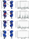
The ribosomal S1 protein (rS1) is indispensable for translation initiation in Gram-negative bacteria. rS1 is a multidomain protein that acts as an RNA chaperone and ensures that mRNAs can bind the ribosome in a single-stranded conformation, which could be related to fast recognition. Although many ribosome structures were solved in recent years, a high-resolution structure of a two-domain mRNA-binding competent rS1 construct is not yet available. Here, we present the NMR solution structure of the minimal mRNA-binding fragment of Vibrio Vulnificus rS1 containing the domains D3 and D4. Both domains are homologues and adapt an oligonucleotide-binding fold (OB fold) motif. NMR titration experiments reveal that recognition of miscellaneous mRNAs occurs via a continuous interaction surface to one side of these structurally linked domains. Using a novel paramagnetic relaxation enhancement (PRE) approach and exploring different spin-labeling positions within RNA, we were able to track the location and determine the orientation of the RNA in the rS1-D34 bound form. Our investigations show that paramagnetically labeled RNAs, spiked into unmodified RNA, can be used as a molecular ruler to provide structural information on protein-RNA complexes. The dynamic interaction occurs on a defined binding groove spanning both domains with identical β2-β3-β5 interfaces. Evidently, the 3'-ends of the cis-acting RNAs are positioned in the direction of the N-terminus of the rS1 protein, thus towards the 30S binding site and adopt a conformation required for translation initiation.
© The Author(s) 2021. Published by Oxford University Press on behalf of Nucleic Acids Research.
Figures


References
-
- Schmeing T.M., Ramakrishnan V.. What recent ribosome structures have revealed about the mechanism of translation. Nature. 2009; 461:1234–1242. - PubMed
-
- Steitz T.A. A structural understanding of the dynamic ribosome machine. Nat. Rev. Mol. Cell Biol. 2008; 9:242–253. - PubMed
-
- Schuwirth B.S., Borovinskaya M.A., Hau C.W., Zhang W., Vila-Sanjurjo A., Holton J.M., Cate J.H.D.. Structures of the bacterial ribosome at 3.5 A resolution. Science. 2005; 310:827–834. - PubMed
-
- Selmer M., Dunham C.M., Murphy F.V, Weixlbaumer A., Petry S., Kelley A.C., Weir J.R., Ramakrishnan V.. Structure of the 70S ribosome complexed with mRNA and tRNA. Science. 2006; 313:1935–1942. - PubMed
-
- Korostelev A., Trakhanov S., Laurberg M., Noller H.F.. Crystal structure of a 70S ribosome-tRNA complex reveals functional interactions and rearrangements. Cell. 2006; 126:1065–1077. - PubMed
Publication types
MeSH terms
Substances
LinkOut - more resources
Full Text Sources
Miscellaneous

