. 2021 Oct;36(5):934-947.
doi: 10.1007/s12250-021-00409-4. Epub 2021 Jul 5.

Antibody Cocktail Exhibits Broad Neutralization Activity Against SARS-CoV-2 and SARS-CoV-2 Variants

Affiliations

Antibody Cocktail Exhibits Broad Neutralization Activity Against SARS-CoV-2 and SARS-CoV-2 Variants

Yuanyuan Qu et al. Virol Sin. 2021 Oct.

Abstract

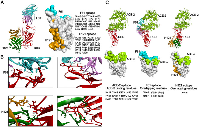
Severe acute respiratory syndrome coronavirus 2 (SARS-CoV-2) has precipitated multiple variants resistant to therapeutic antibodies. In this study, 12 high-affinity antibodies were generated from convalescent donors in early outbreaks using immune antibody phage display libraries. Of them, two RBD-binding antibodies (F61 and H121) showed high-affinity neutralization against SARS-CoV-2, whereas three S2-target antibodies failed to neutralize SARS-CoV-2. Following structure analysis, F61 identified a linear epitope located in residues G446-S494, which overlapped with angiotensin-converting enzyme 2 (ACE2) binding sites, while H121 recognized a conformational epitope located on the side face of RBD, outside from ACE2 binding domain. Hence the cocktail of the two antibodies achieved better performance of neutralization to SARS-CoV-2. Importantly, these two antibodies also showed efficient neutralizing activities to the variants including B.1.1.7 and B.1.351, and reacted with mutations of N501Y, E484K, and L452R, indicated that it may also neutralize the recent India endemic strain B.1.617. The unchanged binding activity of F61 and H121 to RBD with multiple mutations revealed a broad neutralizing activity against variants, which mitigated the risk of viral escape. Our findings revealed the therapeutic basis of cocktail antibodies against constantly emerging SARS-CoV-2 variants and provided promising candidate antibodies to clinical treatment of COVID-19 patients infected with broad SARS-CoV-2 variants.

Keywords: Angiotensin-converting enzyme 2 (ACE2); Antibody cocktail; Broad neutralization; SARS-CoV-2 variants; Severe acute respiratory syndrome coronavirus 2 (SARS-CoV-2).


Conflict of interest statement

The authors declare that they have no conflict of interest.

Figures

Fig. 1
Fig. 1
Generation and screening of antibodies from SARS-CoV-2 convalescent patients. A Antibodies titer in the plasma of SARS-CoV-2 convalescent patients to SARS-CoV-2 N protein and different fragments of SARS-CoV-2 S protein (RBD, S1, and S2). Experiments were performed in duplicate and the error bars denote ± SD, n = 2. B Heat-maps of Fab clones against RBD (n = 288), S1 protein (n = 288) and S2 protein (n = 288). Each lattice represented a Fab clone. C Binding specificity of the 12 candidate IgGs. The binding to different spike proteins (RBD, S1, S2, S protein trimer, and virion) was determined by ELISA Experiments were performed in duplicate, and the error bars denote ± SD, n = 2.
Fig. 2
Fig. 2
Characterizing the binding profile of candidate IgGs. A The specificity of SARS-CoV-2 specific IgGs detected by FACS. HEK 293 T cells expressing SARS-CoV-2 S protein were incubated with candidate mAbs or isotype IgG (HBV mAb) and then stained with anti-human IgG FITC-conjugated antibody. Fluorescence intensity (FITC) negative cells was less than 103, and that of positive cells was around 104. B The affinity of candidate IgGs. The affinity between antibodies (F61, F163, B15, H121, C25, A8, H184, B110, and A199) and S1 was measured by BIAcore 8000 system. Non-competitive ELISA measured the affinity between mAbs (H278, B120, and H285) and S2. C Neutralizing activity of candidate IgGs against SARS-CoV-2 pseudovirus and authentic SARS-CoV-2. Experiments were performed in duplicate, and the error bars denote ± SD, n = 2. The dashed line indicated a 50% reduction in viral infection.
Fig. 3
Fig. 3
Analysis of antibody epitopes by Competition ELISA assays and FACS. A Antigenic epitopes of nine RBD-specific IgGs were analyzed by competitive ELISA. Each lattice shows a competitive percentage. Values less than 0.20 indicated that the antibody has non-competitive epitopes, the value between 0.20 and 0.60 indicated intermediate binding sites, and values greater than 0.60 indicated that the antibody shares overlapping or tight epitopes. B ACE2 binding block assay by FACS. The mouse-Fc tag Fusion protein of SARS-CoV-2 RBD (RBD-mFC) was pre-incubated with nine RBD-specific IgGs or isotype IgG (HBV mAb) and then stained with HEK 293 T cells expressing ACE2. Anti-human (Fc) FITC-conjugated antibody and Anti-mouse (Fc) Texas red-conjugated antibody were used as the secondary antibody. The X-axis represented the fluorescence intensity of human antibodies labeled by FITC, and the Y-axis represented the fluorescence intensity of RBD-mFC labeled by Taxes red.
Fig. 4
Fig. 4
Computer docking (ZDOCK) structure between F61/H121 and SARS-CoV-2 RBD. A ZDOCK structure of the RBD and antibodies complex was shown on the left. RBD was in red. F61 colored blue and pink. H121 was in orange and green. The complex of two antibodies and RBD were superimposed to demonstrate their relative positions and orientations. The footprint of F61 and H121 on RBD was shown in the middle. Blue and orange represent the footprint of F61 and H121, respectively. Binding residues were listed on the right. B The predicted Hydrogen-bond (H-bond) of F61 and H121. Green dashed lines indicated H-bond. The H-bond of F61 was shown on the upper panel. The H-bond of H121 was shown on the lower panel. C Epitopes were overlapping between the two antibodies and ACE2. The interaction between ACE2 and RBD was shown on the left. RBD was in red. ACE2 colored bright green. The interactions between ACE2, RBD and two antibodies were shown on the middle (F61) and left (H121). Color settings were consistent with those mentioned above. Overlapping residues between each of the antibodies and ACE2 were listed at the bottom.
Fig. 5
Fig. 5
Neutralization mutations of SARS-CoV-2 S protein and their effect on antibody neutralizing activity. A Amino acid mutations on S protein. Mutations in B.1.1.7 lineage were labeled red. Mutations in B.1.351 lineage were labeled blue. B The binding activity of RBD-specific IgGs between mutant S1 protein and wild-type (WT) S1 protein was detected by ELISA. The change of binding activity was defined by the ratio of OD450mutant S1/OD450WT S1. The dashed line indicated that the ratio was less than 0.5 or more than 1.5. The significant changes were marked red for decreased. C Neutralization activities of nine RBD-specific IgGs towards mutations on S protein were measured by pseudovirus. D Neutralization activities of F61 and H121 towards mutations K417N, E484K, and N501Y on S protein were measured by pseudovirus. The IC50 value of F61 and H121 were 0.027 μg/mL, 0.078 μg/mL against WT pseudotyped SARS-CoV-2. The changes in neutralization activity were shown in the ratio of IC50 between the variant and WT SARS-CoV-2 (GenBank: MN908947) (IC50WT SARS-CoV-2/IC50 variant SARS-CoV-2). The ratio less than 1 indicated a reduced neutralization activity of the antibody against the variants and the ratio more than 1 indicated an increased neutralization activity. A difference by fourfold is considered significant (ratio more than 4 or less than 0.25). The changes were marked with colored symbols, red for decreased, blue for increased. E Neutralization activities of F61 and H121 towards B1.1.7 and B1.351 were measured by pseudovirus and authentic SARS-CoV-2. The IC50 value of F61 and H121 were 0.027 μg/mL, 0.078 μg/mL against WT pseudotyped SARS-CoV-2 and 0.46 μg/mL, 0.48 μg/mL against WT authentic SARS-CoV-2. The Y-axis represents the fold change of IC50 between the variant and WT SARS-CoV-2 (GenBank: MN908947) (IC50WT SARS-CoV-2/IC50 variant SARS-CoV-2). The ratio less than 1 indicated a reduced neutralization activity of the antibody against the variants and the ratio more than 1 indicated an increased neutralization activity. A difference by fourfold is considered significant (ratio more than 4 or less than 0.25). The red dashed line indicated the ratio was less than 0.25.

References

    1. Barbas CF, Burton DR. Selection and evolution of high-affinity human anti-viral antibodies. Trends Biotechnol. 1996;14:230–234. doi: 10.1016/0167-7799(96)10029-9. - DOI - PubMed
    1. Barbas CF, Kang AS, Lerner RA, Benkovic SJ. Assembly of combinatorial antibody libraries on phage surfaces: the gene III site. Proc Natl Acad Sci U S A. 1991;88:7978–7982. doi: 10.1073/pnas.88.18.7978. - DOI - PMC - PubMed
    1. Barnes CO, Jette CA, Abernathy ME, Dam KA, Esswein SR, Gristick HB, Malyutin AG, Sharaf NG, Huey-Tubman KE, Lee YE, Robbiani DF, Nussenzweig MC, West AP, Jr, Bjorkman PJ. SARS-CoV-2 neutralizing antibody structures inform therapeutic strategies. Nature. 2020;588:682–687. doi: 10.1038/s41586-020-2852-1. - DOI - PMC - PubMed
    1. Benton DJ, Wrobel AG, Xu P, Roustan C, Martin SR, Rosenthal PB, Skehel JJ, Gamblin SJ. Receptor binding and priming of the spike protein of SARS-CoV-2 for membrane fusion. Nature. 2020;588:327–330. doi: 10.1038/s41586-020-2772-0. - DOI - PMC - PubMed
    1. Bertoglio F, Meier D, Langreder N, Steinke S, Rand U, Simonelli L, Heine PA, Ballmann R, Schneider KT, Roth KDR, Ruschig M, Riese P, Eschke K, Kim Y, Schäckermann D, Pedotti M, Kuhn P, Zock-Emmenthal S, Wöhrle J, Kilb N, Herz T, Becker M, Grasshoff M, Wenzel EV, Russo G, Kröger A, Brunotte L, Ludwig S, Fühner V, Krämer SD, Dübel S, Varani L, Roth G, Čičin-Šain L, Schubert M, Hust M. SARS-CoV-2 neutralizing human recombinant antibodies selected from pre-pandemic healthy donors binding at RBD-ACE2 interface. Nature Commun. 2021;12:1577. doi: 10.1038/s41467-021-21609-2. - DOI - PMC - PubMed

Substances