. 2022 Jan;237(1):551-565.
doi: 10.1002/jcp.30501. Epub 2021 Jul 5.

Lipocalin 2 increases after high-intensity exercise in humans and influences muscle gene expression and differentiation in mice

Affiliations

Lipocalin 2 increases after high-intensity exercise in humans and influences muscle gene expression and differentiation in mice

Marco Ponzetti et al. J Cell Physiol. 2022 Jan.

Abstract

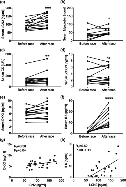
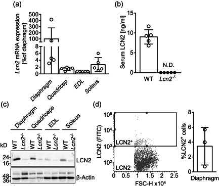
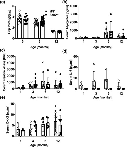
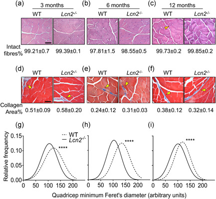
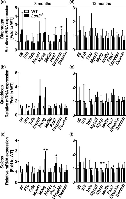
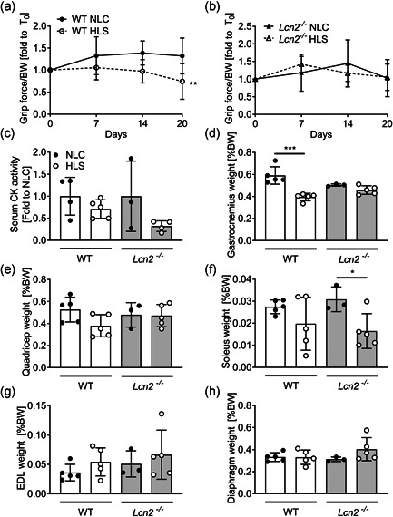
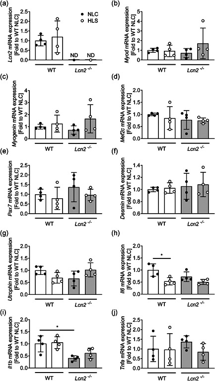
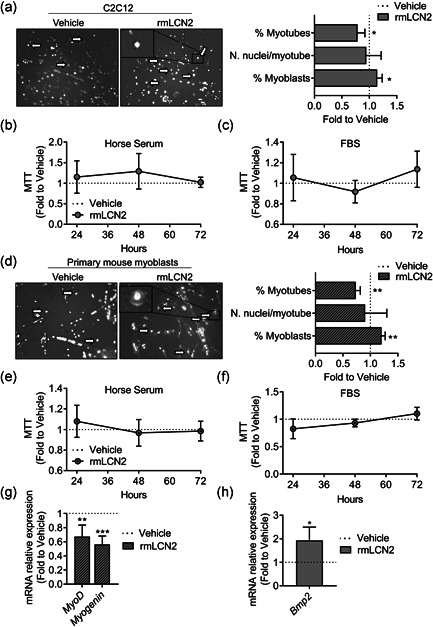
Lipocalin 2 (LCN2) is an adipokine that accomplishes several functions in diverse organs. However, its importance in muscle and physical exercise is currently unknown. We observed that following acute high-intensity exercise ("Gran Sasso d'Italia" vertical run), LCN2 serum levels were increased. The Wnt pathway antagonist, DKK1, was also increased after the run, positively correlating with LCN2, and the same was found for the cytokine Interleukin 6. We, therefore, investigated the involvement of LCN2 in muscle physiology employing an Lcn2 global knockout (Lcn2-/- ) mouse model. Lcn2-/- mice presented with smaller muscle fibres but normal muscle performance (grip strength metre) and muscle weight. At variance with wild type (WT) mice, the inflammatory cytokine Interleukin 6 was undetectable in Lcn2-/- mice at all ages. Intriguingly, Lcn2-/- mice did not lose gastrocnemius and quadriceps muscle mass and muscle performance following hindlimb suspension, while at variance with WT, they lose soleus muscle mass. In vitro, LCN2 treatment reduced the myogenic differentiation of C2C12 and primary mouse myoblasts and influenced their gene expression. Treating myoblasts with LCN2 reduced myogenesis, suggesting that LCN2 may negatively affect muscle physiology when upregulated following high-intensity exercise.

Keywords: adipokines; bone; exercise; lipocalin 2; muscle; myogenesis.


Figures

Figure 1
Figure 1
“Gran Sasso d'Italia” vertical run. Fifteen participants took part in the vertical run. Blood was collected right before and after the race. (a–f) Serum analyses showing levels of (a) LCN2, (b) myoglobin, (c) creatine kinase (CK), (d) undercarboxylated osteocalcin (ucOCN), (E) DKK1 and (F) IL6. (G,H) Pearson's correlation analyses between LCN2 and (g) DKK1 or (h) IL6 serum levels. (a–f) Paired Student's t test. *p < .05, **p < .01, ***p < .001 and ****p < .0001 after race vs before race
Figure 2
Figure 2
LCN2 expression in mouse muscle tissues. (a) Lcn2 mRNA expression analysed in muscle sections from diaphragm, quadriceps, EDL and soleus of WT male mice. Results are the % to the average of the diaphragm ΔC ts (N = 5 mice per group). (b) ELISA showing circulating levels of LCN2 in WT and Lcn2 −/− mice. (c) Western blot representative of three mice per group, showing muscle expression of LCN2 and β‐Actin as a loading control. (d) Cytofluorimetric analysis of mononuclear cells extracted from the diaphragm of 6‐month‐old WT mice and stained for LCN2. Left panel: representative dot plot with gating box; right panel: quantification of percent LCN2 positive cells. EDL, Extensor Digitorum Longus; ELISA, enzyme‐linked immunosorbent assay; mRNA, messenger RNA; WT, wild‐type
Figure 3
Figure 3
Effect of the lack of LCN2 on muscle phenotype. WT and Lcn2 ‐/− mice were subjected to (a) grip force analysis at the ages indicated in the abscissa. Results are expressed as grip force (g)/body weight(g). After sacrifice, sera were harvested and analysed for (b) myoglobin, (c) CK, (d) IL6 and (b, d, e) DKK1 by ELISA or (c) by the Reflotron assay. Curve fitting test. *p < .05 versus WT. CK, creatine kinase; ELISA, enzyme‐linked immunosorbent assay; IL6, interleukin‐6; WT, wild‐type
Figure 4
Figure 4
Histological analysis of WT andLcn2 −/− mice quadriceps. Quadriceps from WT and Lcn2 −/− mice were explanted, fixed, paraffin‐embedded and sectioned at 5 μm. (a–c) Haematoxylin‐eosin staining performed on histological sections from (a) 3‐, (b) 6‐, and (c) 12‐month‐old mice to quantify the % of intact fibres. (d–f) Masson's trichrome staining to evaluate the % collagen area on histological sections from (d) 3‐, (e) 6‐ and (f) 12‐month‐old mice. (g–i) After haematoxylin‐eosin staining, minimum Feret's diameter from quadricep fibres was assessed via software (NIH ImageJ, version 1.50i.) in 3‐, (h) 6‐, and (i) 12‐month‐old mice. Gaussian curves were interpolated to better represent the fibre size distributions. (a–f) N = 3–5 mice per group; (g–i) N > 1000 fibres per group, arising from N = 3–5 mice per group. Scale bar = 50 μm; orange arrowhead: example of non‐intact (centrally nucleated) fibre; yellow arrowhead: collagen area stained in blue. Student's t test. ****p < .0001 versus WT. WT, wild‐type
Figure 5
Figure 5
Transcriptional profiling of WT andLcn2 −/−mice muscles. RNA was extracted from (a–c) 3‐ or (d–f) 12‐month‐old WT and Lcn2 −/− mice muscles. One μg of total RNA was reverse‐transcribed into cDNA and used for transcriptional analyses. (a, d) Diaphragm, (b, e) quadriceps and (c, f) soleus. The genes analysed are indicated in the abscissa. N = 5–10. Student's t test or Mann–Whitney test. *p < .05; **p < .01 vs WT. cDNA, complmentary DNA; IL6, interleukin‐6; mRNA, messenger RNA; WT, wild‐type
Figure 6
Figure 6
Effect of the lack of Lcn2 on muscle phenotype under mechanical unloading conditions. Eight‐week‐old WT and Lcn2 −/− mice were subjected to hindlimb suspension for 3 weeks. (a, b) Grip force was performed during the timeframe of the experiment in hindlimb suspended (HLS) or normal loading condition (NLC) (a) WT and (b)Lcn2 −/− mice. At sacrifice, serum was harvested to measure (c) CK serum activity. (d) Gastrocnemius, (e) quadriceps, (d) soleus, (g) EDL and (H) diaphragm muscles explanted from HLS and NLC mice were weighted and plotted as % of body weight. (a, b) Curve fitting test, N = 3–5. (c–h) Student's t test. *p < .05; ***p < .01; ***p < .001. CK, creatine kinase; EDL, Extensor Digitorum Longus; WT, wild‐type
Figure 7
Figure 7
Effect of the lack of Lcn2 on muscle gene expression under mechanical unloading conditions. Eight‐week‐old WT and Lcn2 −/− mice were subjected to hindlimb suspension for 3 weeks. At sacrifice, quadriceps RNA was extracted, reverse‐transcribed and subjected to real‐time RT‐PCR to analyse the expression of (a) Lcn2, (b) Myod, (c) Myogenin, (d) Mef2C, (e) Pax7, (f) Desmin, (g) Utrophin, (H) Il6, (I) Il1b, and (J) Tnfa. Results are shown as fold to WT NLC. Student's t test. *p < .05. IL, interleukin; RT‐PCR, reverse transcrition polrmerase chain reaction
Figure 8
Figure 8
Effect of rmLCN2 on muscle cells in vitro. (a) C2C12 cells were treated with 1 µg/ml LCN2 under a myogenic differentiation medium (DMEM supplemented with 5% horse serum) for 4 days. Then cells were fixed and stained with DAPI to evidence nuclei. Merged phase‐contrast DAPI images were collected to analyse the % of myonuclei, the % of myoblasts and the number of nuclei/myotube. (b) C2C12 were treated with 1 µg/ml rmLCN2 for 24, 48, or 72 h under differentiation medium or (c) DMEM plus 10% FBS. MTT was carried out to analyse cell metabolic activity. (d) Primary CD1 mouse myoblasts were treated with 200 ng/ml rmLCN2 for 24, 48, or 72 h under differentiation medium, then cells were fixed and stained with DAPI to evidence nuclei. Merged phase‐contrast DAPI images were collected to analyse the % of myonuclei, the % of myoblasts and the number of nuclei/myotube. (e) Myoblasts were treated with 200 ng/ml rmLCN2 for 24, 48, or 72 h under differentiation medium or (f) DMEM/F10 plus 20% FBS and 2.5 ng/ml bFGF. MTT was carried out to analyse cell metabolic activity. (g) Myoblasts were treated for 4 days with 200 ng/ml rmLCN2 under differentiation medium, then RNA was extracted, reverse‐transcribed, and Myod and Myogenin expression was analysed. (h) C2C12 cells were treated as described in (a), then RNA was extracted, reverse‐transcribed and subjected to signal transduction pathway finder RT2‐array. After applying cut‐off criteria (threshold cycle [C t]<30, p‐value <.05) and validating the results with a separate set of primer, we found Bmp2 to be significantly upregulated in cells treated with rmLCN2. (a, d) Arrow and insets: myoblast‐like cells. Student's t test. *p < .05; **p < .01 versus vehicle. DMEM, Dulbecco's modified Eagle's medium; bFGF, basic fibroblast growth factor; DAPI, 4′,6‐diamidino‐2‐phenylindole; FBS, fetal bovine serum; MTT, 3‐(4,5‐Dimethylthiazol‐2‐yl)‐2,5‐diphenyltetrazolium

References

    1. Abella, V. , Scotece, M. , Conde, J. , Gómez, R. , Lois, A. , Pino, J. , Gómez‐Reino, J. J. , Lago, F. , Mobasheri, A. , & Gualillo, O. (2015). The potential of lipocalin‐2/NGAL as biomarker for inflammatory and metabolic diseases. Biomarkers, 20(8), 565–571. 10.3109/1354750X.2015.1123354 - DOI - PMC - PubMed
    1. Barclay, C. J. , Constable, J. K. , & Gibbs, C. L. (1993). Energetics of fast‐ and slow‐twitch muscles of the mouse. The Journal of Physiology, 472, 61–80. 10.1113/jphysiol.1993.sp019937 - DOI - PMC - PubMed
    1. Berger, T. , Togawa, A. , Duncan, G. S. , Elia, A. J. , You‐Ten, A. , Wakeham, A. , Fong, H. E. , Cheung, C. C. , & Mak, T. W. (2006). Lipocalin 2‐deficient mice exhibit increased sensitivity to Escherichia coli infection but not to ischemia‐reperfusion injury. Proceedings of the National Academy of Sciences of the United States of America, 103(6), 1834–1839. 10.1073/pnas.0510847103 - DOI - PMC - PubMed
    1. Burattini, S. , Ferri, R. , Battistelli, M. , Curci, R. , Luchetti, F. , Falcieri, E. (2004). C2C12 murine myoblasts as a model of skeletal muscle development: Morpho‐functional characterization, European Journal of Histochemistry, 48(3), 223–233, 10.4081/891 - DOI - PubMed
    1. Capulli, M. , Ponzetti, M. , Maurizi, A. , Gemini‐Piperni, S. , Berger, T. , Mak, T. W. , Teti, A. , & Rucci, N. (2018). A Complex role for Lipocalin 2 in bone metabolism: Global ablation in mice induces osteopenia caused by an altered energy metabolism. Journal of Bone and Mineral Research, 33(6), 1141–1153. 10.1002/jbmr.3406 - DOI - PubMed

Publication types

Grants and funding

LinkOut - more resources