Review
. 2022 Feb;35(1):3-24.
doi: 10.1177/19714009211029177. Epub 2021 Jul 5.

A review of neuroradiological abnormalities in patients with coronavirus disease 2019 (COVID-19)

Affiliations
Review

A review of neuroradiological abnormalities in patients with coronavirus disease 2019 (COVID-19)

Bahar Bahranifard et al. Neuroradiol J. 2022 Feb.

Abstract

Background: Severe acute respiratory syndrome coronavirus 2 (SARS-CoV-2) has led to various neurological manifestations. There is an urgent need for a summary of neuroimaging findings to accelerate diagnosis and treatment plans. We reviewed prospective and retrospective studies to classify neurological abnormalities observed in patients with the SARS-CoV-2 infection.

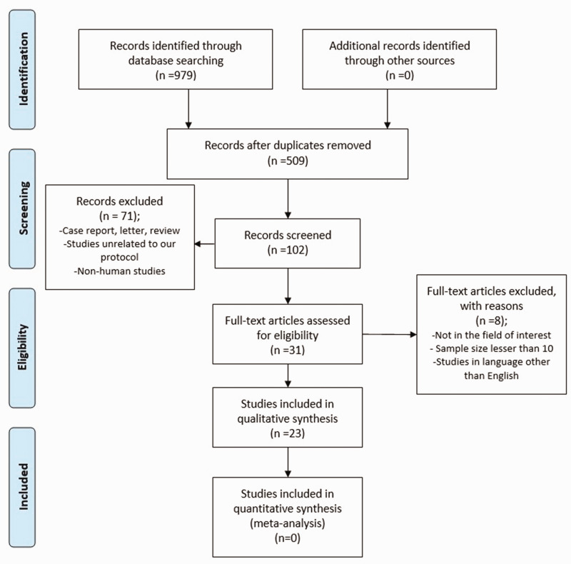
Methods: The relevant studies published in Scopus, PubMed and Clarivate Analytics databases were analysed. The search was performed for full-text articles published from 23 January 2020 to 23 February 2021.

Results: In 23 studies the number of patients with SARS-CoV-2 infection was 20,850 and the number of patients with neurological manifestations was 1996 (9.5%). The total number of patients with neuroradiological abnormalities was 602 (2.8%). SARS-CoV-2 has led to various neuroimaging abnormalities which can be categorised by neuroanatomical localisation of lesions and their main probable underlying pathogenesis. Cranial nerve and spinal root abnormalities were cranial neuritis and polyradiculitis. Parenchymal abnormalities fell into four groups of: (a) thrombosis disorders, namely ischaemic stroke and sinus venous thrombosis; (b) endothelial dysfunction and damage disorders manifested as various types of intracranial haemorrhage and posterior reversible encephalopathy syndrome; (c) hypoxia/hypoperfusion disorders of leukoencephalopathy and watershed infarction; and (d) inflammatory disorders encompassing demyelinating disorders, encephalitis, vasculitis-like disorders, vasculopathy and cytotoxic lesions of the corpus callosum. Leptomeninges disorders included meningitis. Ischaemic stroke was the most frequent abnormality in these studies.

Conclusion: The review study suggests that an anatomical approach to the classification of heterogeneous neuroimaging findings in patients with SARS-CoV-2 and neurological manifestations would lend itself well for use by practitioners in diagnosis and treatment planning.

Keywords: COVID-19; Neuroradiological; brain; computed tomography; magnetic resonance; spinal.


Figures

Figure 1.
Figure 1.
The preferred reporting items for systematic reviews and meta-analysis (PRISMA) flow diagram.
Figure 2.
Figure 2.
Pathophysiological mechanisms of neuroradiological abnormalities of patients with severe acute respiratory syndrome coronavirus 2 (SARS-CoV-2) infection. ANE: acute necrotising encephalopathy; CLOCC: cytotoxic lesions of the corpus callosum; CLE: cerebral leukoencephalopathy; HIP/ARS: hyperinflammatory phase/acute respiratory phase; MH: microhaemorrhage; PRES: posterior reversible encephalopathy syndrome; SVT: sinus venous thrombosis.
Figure 3.
Figure 3.
Neuroradiological abnormalities of patients with severe acute respiratory syndrome coronavirus 2 (SARS-CoV-2) infection. ADEM: acute disseminated encephalomyelitis; CLOCC: cytotoxic lesions of the corpus callosum; CLE: cerebral leukoencephalopathy; MH: microhaemorrhage; PRES: posterior reversible encephalopathy syndrome; SVT: sinus venous thrombosis.
Figure 4.
Figure 4.
(a)–(d) Facial and vestibular neuritis. (a) and (b) A woman in her early 60s with severe acute respiratory syndrome coronavirus 2 (SARS-CoV-2) infection and facial neuritis. Precontrast (a) and postcontrast (b) T1-weighted imaging (WI) brain magnetic resonance imaging (MRI) showed bilateral facial nerve enhancement (b, arrow). (c) and (d) A woman in her mid-40s with SARS-CoV-2 infection and right-sided vestibular neuritis. Precontrast (c) and postcontrast (d) T1-WI brain MRI showed enhancement of the right vestibular nerve (d, arrow). (e)–(h) Polyradiculitis in a woman in her mid-30s with SARS-CoV-2 infection, lumbar pain and leg paresthesia. (e) and (f) Precontrast (e) and postcontrast (f) sagittal lumbar T1-WI showed faint contrast enhancement of the cauda equina (f, arrow). (g) and (h) Precontrast (g) and postcontrast (h) axial T1-WI images showed faint enhancement of multiple nerve roots (h, arrow).
Figure 5.
Figure 5.
Posterior reversible encephalopathy syndrome in a 66-year-old man with severe acute respiratory syndrome coronavirus 2 (SARS-CoV-2) and seizure. Fluid-attenuated inversion recovery (FLAIR) sequence showed cortical and subcortical hyperintensity in the occipital lobe extending to parietal and frontal lobes (a) and (d, arrow), without restriction on diffusion-weighted imaging (DWI) (b) and (e). The follow-up magnetic resonance imaging (MRI) indicated the complete disappearance of the lesions (c) and (f).
Figure 6.
Figure 6.
Leukoencephalopathy with diffuse microhaemorrhage in an elderly man with severe acute respiratory syndrome coronavirus 2 (SARS-CoV-2) infection and delay in awakening after sedation withdrawal. Brain magnetic resonance imaging (MRI) shows diffuse leukoencephalopathy with the involvement of subcortical on fluid-attenuated inversion recovery (FLAIR) (a) and (b) and diffuse microhaemorrhage with a predilection for subcortical U-fibres and the corpus callosum on susceptibility weighted imaging (SWI) (c).
Figure 7.
Figure 7.
Acute haemorrhagic encephalomyelitis in a 52-year-old man with severe acute respiratory syndrome coronavirus 2 (SARS-CoV-2) infection. Initial T2 weighted imaging (WI) showed confluent multifocal hyperintense lesions in the white matter, corpus callosum, internal and external capsulesonT2-WI (a) and (b), with progression in the follow-up study, ending in diffusely involved white matter (d) and (e). Susceptibility weighted imaging (SWI) showed microhaemorrhages changes (c) and (f, arrow) and prominent medullary veins (f, short arrows).
Figure 8.
Figure 8.
Temporal lobe encephalitis in a 58-year-old man with severe acute respiratory syndrome coronavirus 2 (SARS-CoV-2) infection. Fluid-attenuated inversion recovery (FLAIR) sequence showed hyperintensities (arrow) in the left medial temporal lobe.
Figure 9.
Figure 9.
Acute necrotising encephalitis in a 59-year-old woman with severe acute respiratory syndrome coronavirus 2 (SARS-CoV-2) infection. T2-weighted image (WI)sequence showed extensive hyper signal lesions of the bilateral medial temporal lobes, basal ganglia, thalami and pons (a), and a restriction on diffusion-weighted imaging (DWI) (c) with punctate haemorrhage change on susceptibility-weighted imaging (SWI) (b) and enhancement on post-contrast T1-WI (d, arrow).
Figure 10.
Figure 10.
Encephalitis with nigrostriatal pathway involvement in a 42-year-old man with chronic lymphocytic leukaemia and severe acute respiratory syndrome coronavirus 2 (SARS-CoV-2) infection. Brain magnetic resonance imaging (MRI) shows a diffusion restriction of the substantia nigra on diffusion-weighted imaging (DWI) (a, arrows) with corresponding decreased signal on apparent diffusion coefficient (ADC) (b, arrows) and hyperintensity on fluid-attenuated inversion recovery (FLAIR) (c, arrows).
Figure 11.
Figure 11.
Cytotoxic lesion of the corpus callosum (CLOCC) in a woman in her late 40s with severe acute respiratory syndrome coronavirus 2 (SARS-CoV-2). It showed a hypersignal lesion in the posterior portion of the corpus callosum on T2-weighted image (WI) and fluid-attenuated inversion recovery (FLAIR) (c) and (d) with restriction on diffusion-weighted imaging (DWI) (a) and corresponding decreased signal on apparent diffusion coefficient (ADC) (b).
Figure 12.
Figure 12.
Cerebral vasculitis-like lesion in a 50-year-old man with severe acute respiratory syndrome coronavirus 2 (SARS-CoV-2) infection. Brain magnetic resonance imaging (MRI) shows multiple punctiform and oedematous lesions in the periventricular white matter which are hypersignal on fluid-attenuated inversion recovery (FLAIR) (a) and (e, arrow), a diffusion restriction on the diffusion-weighted imaging (DWI) (b) and (f, arrow) without decreased apparent diffusion coefficient (ADC) (c) and (g, arrow), and a perivascular enhancement on the post-contrast T1-weighted image (WI) (d) and (h, arrow).
Figure 13.
Figure 13.
Vasculopathy (arterial narrowing) and stroke in a 54-year-old male patient with severe acute respiratory syndrome coronavirus 2 (SARS-CoV-2) infection. Non-contrast computed tomography (CT) of the head showed initial temporal haemorrhage (a, arrow) with surrounding parenchyma oedema. Magnetic resonance angiography (MRA) showed focal stenosis of the supraclinoid internal carotid artery (b, arrow) with a patent lumen and flow distal to stenosis (b, double arrows). Diffusion-weighted imaging (DWI) revealed a restriction in the right middle cerebral artery (MCA) territory, suggestive of acute cerebral infarction (c) and (d, arrow).
Figure 14.
Figure 14.
Vasculopathy (arterial narrowing and vessel wall enhancement) in a 69-year-old male patient with severe acute respiratory syndrome coronavirus 2 (SARS-CoV-2) infection and delay in awakening after sedative withdrawal. (a)–(f) Precontrast T1-weighted image (WI) was normal, but post-contrast T1-WI showed vessels wall enhancement of the basilar artery (b) and (c), the left middle cerebral artery (d) and the right posterior cerebral artery (PCA) (e), as well as narrowing of both PCAs on brain magnetic resonance angiography (MRA) (f).
Figure 15.
Figure 15.
Frequency of neuroradiological abnormalities in 602 patients with severe acute respiratory syndrome coronavirus 2 (SARS-CoV-2) infection.

References

    1. Mao L, Jin H, Wang M, et al. Neurologic manifestations of hospitalized patients with coronavirus disease 2019 in Wuhan, China. JAMA Neurology 2020; 77: 683–690. - PMC - PubMed
    1. Somani S, Agnihotri SP. Emerging neurology of COVID-19. The Neurohospitalist 2020; 10: 281–286. - PMC - PubMed
    1. Ellul MA, Benjamin L, Singh B, et al. Neurological associations of COVID-19. Lancet Neurol 2020; 19: 767–783. - PMC - PubMed
    1. Virhammar J, Kumlien E, Fällmar D, et al. Acute necrotizing encephalopathy with SARS-CoV-2 RNA confirmed in cerebrospinal fluid. Neurology 2020; 95: 445. - PMC - PubMed
    1. Afshar H, Yassin Z, Kalantari S, et al. Evolution and resolution of brain involvement associated with SARS-CoV2 infection: a close clinical-paraclinical follow up study of a case. Mult Scler Relat Disord 2020; 43: 102216–102216. 2020/05/21. - PMC - PubMed