. 2021 Sep 15:339:235-242.
doi: 10.1016/j.ijcard.2021.06.056. Epub 2021 Jul 3.

Cardiac involvement in consecutive unselected hospitalized COVID-19 population: In-hospital evaluation and one-year follow-up

Collaborators, Affiliations

Cardiac involvement in consecutive unselected hospitalized COVID-19 population: In-hospital evaluation and one-year follow-up

Viviana Maestrini et al. Int J Cardiol. .

Abstract

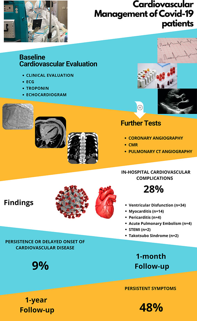
Background: Cardiovascular disease (CVD) can occur in COVID-19 and has impact on clinical course. Data on CVD prevalence in hospitalized COVID-19 patients and sequelae in survivors is limited. Aim of this prospective study carried out on consecutive unselected COVID-19 population, was to assess: 1) CVD occurrence among hospitalized COVID-19 patients, 2) persistence or new onset of CVD at one-month and one-year follow-up.

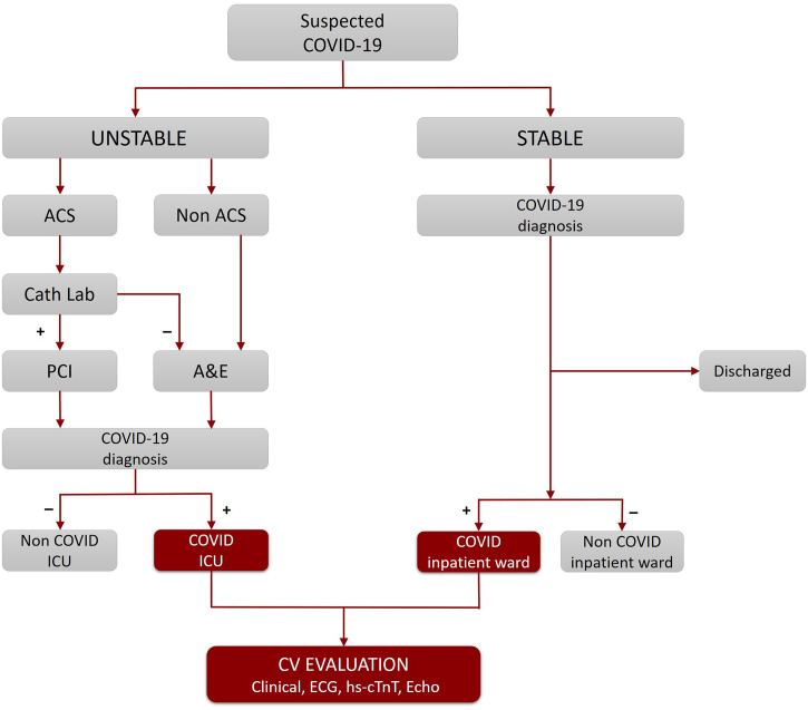
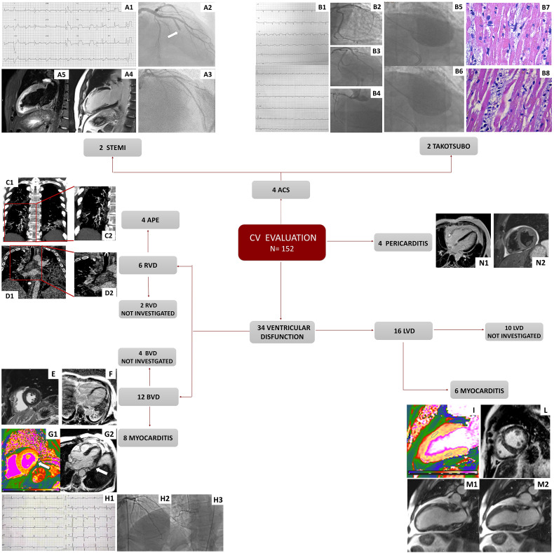
Methods: Over 30 days n = 152 COVID-19 patients underwent cardiovascular evaluation. Standard electrocardiogram (ECG), Troponin and echocardiography were integrated by further tests when indicated. Medical history, arterial blood gas, blood tests, chest computed tomography and treatment were recorded. CVD was defined as the occurrence of a new condition during the hospitalization for COVID-19. Survivors attended a one-month follow-up visit and a one-year telephone follow-up.

Results: Forty-two patients (28%) experienced a wide spectrum of CVD with acute myocarditis being the most frequent. Death occurred in 32 patients (21%) and more frequently in patients who developed CVD (p = 0.032). After adjustment for confounders, CVD was independently associated with death occurrence. At one-month follow-up visit, 7 patients (9%) presented persistent or delayed CVD. At one-year telephone follow-up, 57 patients (48%) reported persistent symptoms.

Conclusion: Cardiovascular evaluation in COVID-19 patients is crucial since the occurrence of CVD in hospitalized COVID-19 patients is common (28%), requires specific treatment and increases the risk of in-hospital mortality. Persistence or delayed presentation of CVD at 1-month (9%) and persistent symptoms at 1-year follow-up (48%) suggest the need for monitoring COVID-19 survivors.

Keywords: COVID-19; Cardiovascular disease; Cardiovascular magnetic resonance; Echocardiography; Follow-up; Myocarditis.


Conflict of interest statement

The authors report no relationships that could be construed as a conflict of interest.

Figures

Unlabelled Image
Graphical abstract
Fig. 1
Fig. 1
Recruitment flow chart. This flow chart illustrates the patient care pathway from A&E admission to hospital ward assignment. A&E: Accident & Emergency; ACS: Acute Coronary Syndrome; CV: Cardiovascular; hs-cTnT: high-sensitivity cardiac troponin T; ICU: Intensive Care Unit; PCI: Percutaneous Coronary Intervention.
Fig. 2
Fig. 2
Examples of cardiac events. The figure shows examples of in-hospital CVD. Fig. A: patient presented with anterior STEMI (A1), with obstructed LAD (A2-A3), oedema (A4) and LGE (A5) at the mid-apical anterior segments by CMR (A4-A5). Fig. B: patient presented with diffuse ST segment elevation (B1), unobstructed coronary arteries (B2-B4) and mid-apical ballooning at the ventricular angiography (end-diastolic frame, B5, and end-systolic frame, B6). Autopsy revealed presence of contraction band in cardiomyocytes (B7) and marked interstitial oedema and mononucleate cells, in absence of myocyte necrosis (B8). Fig. C1 (magnification in C2): patient with thrombi in the segmental pulmonary arterial branches for the posterior and middle-basal segment of the right lower lobe while in D1 (magnification in D2) a patient with intraluminal thrombus in the arterial branch for the lower right lobe. Figs. E-G: three cases of CMR findings suggestive of myocarditis: non-ischemic LGE (sub-epicardial at the inferior wall and mid-myocardial at the inferior septum) (E); sub-epicardial LGE areas at the lateral wall (breathing artefact in the image) (F); short axis high native T1 Mapping at the basal lateral wall (G1) and sub-epicardial LGE at the same level (G2) in 3 chamber view. Fig. H: patient presenting with severe biventricular dysfunction, troponin raise, diffuse ECG changes (H1) and unobstructed coronary arteries (H2−H3). Fig. I: patient with myocarditis with high native T1 Mapping (ROI in the anterior wall: 1133 ms, in the inferior wall: 1030 ms). Fig. L: non-ischemic LGE at the inferior RV insertion point. Multiple areas of increased signal in the cine (M1 end-diastolic frame and M2 end-systolic frame) sequences at the subpericardial level in the anterior and inferior wall. Patient with acute pericarditis (N1−N2).

Comment in

References

    1. Wang D., Hu B., Hu C., et al. Clinical characteristics of 138 hospitalized patients with 2019 novel coronavirus-infected pneumonia in Wuhan, China. Jama. 2020;323:1061–1069. - PMC - PubMed
    1. Huang C., Wang Y., Li X., et al. Clinical features of patients infected with 2019 novel coronavirus in Wuhan, China. Lancet. 2020;395:497–506. - PMC - PubMed
    1. Guo T., Fan Y., Chen M., et al. Cardiovascular implications of fatal outcomes of patients with coronavirus disease 2019. JAMA Cardiol. 2020;5:811–818. - PMC - PubMed
    1. Meyer P., Degrauwe S., Van Delden C., Ghadri J.R., Templin C. Typical takotsubo syndrome triggered by SARS-CoV-2 infection. Eur. Heart J. 2020;41:1860. - PMC - PubMed
    1. Bangalore S., Sharma A., Slotwiner A., et al. ST-segment elevation in patients with Covid-19 - a case series. N. Engl. J. Med. 2020;382:2478–2480. - PMC - PubMed