Poroid hidradenoma of the scalp
- PMID: 34225407
- PMCID: PMC8257446
- DOI: 10.7181/acfs.2021.00101
Poroid hidradenoma of the scalp
Abstract
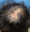
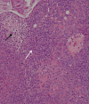
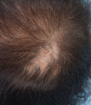
Poroid hidradenoma has both features of hidradenoma and poroma. The histological hidradenoma framework consisting of solid and cystic components, and the presence of poroid and cuticular cells resembling a poroid neoplasm. Despite transforming into malignant neoplasm only in < 1% of cases, its histological characteristics may resemble those of malignant neoplasms. Although the risk of malignant transformation is very low, surgical excision is recommended to prevent growth and/or recurrence. To date, very few cases of poroid hidradenoma have been reported in the literature. Herein, we present a case of poroid hidradenoma on the scalp of a 74-year-old woman.
Keywords: Eccrine glands; Poroma; Skin neoplasms.
Conflict of interest statement
No potential conflict of interest relevant to this article was reported.
Figures


References
-
- Abenoza P, Ackerman AB. Poromas. In: Abenoza P, Ackerman AB, editors. Neoplasms with eccrine differentiation. Philadelphia: Lea & Febiger; 1990. p. 113.
-
- Ito K, Ansai SI, Fukumoto T, Anan T, Kimura T. Clinicopathological analysis of 384 cases of poroid neoplasms including 98 cases of apocrine type cases. J Dermatol. 2017;44:327–34. - PubMed
-
- Requena L, Sanchez M. Poroid hidradenoma: a light microscopic and immunohistochemical study. Cutis. 1992;50:43–6. - PubMed
-
- Penneys NS, Ackerman AB, Indgin SN, Mandy SH. Eccrine poroma: two unusual variants. Br J Dermatol. 1970;82:613–5. - PubMed
Publication types
LinkOut - more resources
Full Text Sources

