Evaluation of the optimal dosage of BMP-9 through the comparison of bone regeneration induced by BMP-9 versus BMP-2 using an injectable microparticle embedded thermosensitive polymeric carrier in a rat cranial defect model
- PMID: 34225891
- PMCID: PMC8260961
- DOI: 10.1016/j.msec.2021.112252
Evaluation of the optimal dosage of BMP-9 through the comparison of bone regeneration induced by BMP-9 versus BMP-2 using an injectable microparticle embedded thermosensitive polymeric carrier in a rat cranial defect model
Abstract
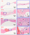
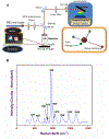
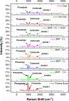
Bone morphogenetic proteins (BMPs) are well known as enhancers and facilitators of osteogenesis during bone regeneration. The use of recombinant BMP-2 (rhBMP-2) in bone defect healing has drawbacks, which has driven the scouting for alternatives, such as recombinant BMP-9 (rhBMP-9), to provide comparable new bone formation. However, the dosage of rhBMP-9 is quintessential for the facilitation of adequate bone defect healing. Therefore, this study has been designed to evaluate the optimal dosage of BMP-9 by comparing the bone defect healing induced by rhBMP-9 over rhBMP-2. The chitosan (CS) microparticles (MPs), coated with BMPs, were embedded in a thermoresponsive methylcellulose (MC) and calcium alginate (Alg) based injectable delivery system containing a dosage of either 0.5 μg or 1.5 μg of the respective rhBMP per bone defect. A 5 mm critical-sized cranial defect rat model has been used in this study, and bone tissues were harvested at eight weeks post-surgery. The standard tools for comparing the new bone regeneration included micro computerized tomography (micro-CT) and histological analysis. A novel perspective of analyzing the new bone quality and crystallinity was employed by using Raman spectroscopy, along with its elastic modulus quantified through Atomic Force Microscopy (AFM). Results showed that the rhBMP-9 administered at a dosage of 1.5 μg per bone defect, using this delivery system, can adequately facilitate the bone void filling with ample new bone mineralization and crystallinity as compared to rhBMP-2, thus approving the hypothesis for a viable rhBMP-2 alternative.
Keywords: Atomic force microscopy; Bone morphogenetic protein; Bone regeneration; Microparticles; Raman spectroscopy.
Copyright © 2021 Elsevier B.V. All rights reserved.
Conflict of interest statement
Conflict of Interest
The authors would like to state that there are no conflicts of interest.
Declaration of interests
The authors declare that they have no known competing financial interests or personal relationships that could have appeared to influence the work reported in this paper.
Figures


References
-
- Gautschi OP, Frey SP, Zellweger R, ANZ J Surg, 77 (2007) 626–631. - PubMed
-
- Carreira AC, Lojudice FH, Halcsik E, Navarro RD, Sogayar MC, Granjeiro JM, J. Dent. Res, 93 (2014) 335–345. - PubMed
-
- Tumialan LM, Pan J, Rodts GE, Mummaneni PV, J. Neurosurg, 8 (2008) 529–535. - PubMed
-
- Shields LBE, Raque GH, Glassman SD, Campbell M, Vitaz T, Harpring J, Shields CB, Spine, 31 (2006) 542–547. - PubMed
MeSH terms
Substances
Grants and funding
LinkOut - more resources
Full Text Sources
Miscellaneous

