. 2021:164:167-185.
doi: 10.1016/bs.mcb.2020.10.009. Epub 2020 Nov 18.

Assessment of mammalian endosomal microautophagy

Affiliations

Assessment of mammalian endosomal microautophagy

Gregory J Krause et al. Methods Cell Biol. 2021.

Abstract

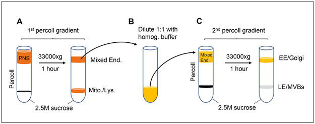
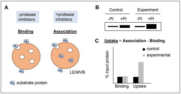
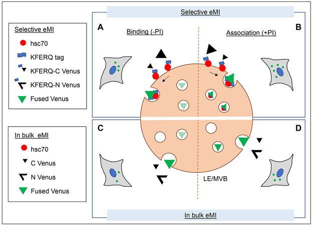
Endosomal microautophagy (eMI) is a type of autophagy that allows for the selective uptake and degradation of cytosolic proteins in late endosome/multi-vesicular bodies (LE/MVB). This process starts with the recognition of a pentapeptide amino acid KFERQ-like targeting motif in the substrate protein by the hsc70 chaperone, which then enables binding and subsequent uptake of the protein into the LE/MVB compartment. The recognition of a KFERQ-like motif by hsc70 is the same initial step in chaperone-mediated autophagy (CMA), a form of selective autophagy that degrades the hsc70-targeted proteins in lysosomes in a LAMP-2A dependent manner. The shared step of substrate recognition by hsc70, originally identified for CMA, makes it now necessary to differentiate between the two pathways. Here, we detail biochemical and imaging-based methods to track eMI activity in vitro with isolated LE/MVBs and in cells in culture using fluorescent reporters and highlight approaches to distinguish whether a protein is a substrate of eMI or CMA.

Keywords: Autophagy; Chaperones; Late endosomes; Multi-vesicular bodies; Organelle isolation; Protein degradation; Protein targeting; Proteostasis.


Figures

Figure 1:
Figure 1:. Scheme of late endosome/multi-vesicular bodies (LE/MVBs) isolation.
Isolation of LE/MVB is attained after tissue homogenization by differential centrifugation to obtain a post-nuclear supernatant (PNS) that is further fractionated by sequential centrifugation through discontinuous density Percoll® and sucrose gradients as depicted. Briefly, the PNS is fist layered on top of a 2.5M sucrose cushion and 27% Percoll® layer gradient and centrifuged to eliminate mitochondria and lysosomes through the Percoll® layer (A). The remaining organelles collected at the top of the gradient are then diluted in homogenization buffer (B) and layered on top of a second 2.5M sucrose cushion/10% Percoll® layer gradient. Upon centrifugation, LE/MVB migrate through the 10% Percoll® layer and can be collected at the interface of the sucrose/Percoll® layers, whereas early endosomes (EE) and Golgi fractions remain at the top of the gradient (C).
Figure 2:
Figure 2:. in vitro reconstitution of eMI with isolated LE/MVBs.
A. Scheme of the reconstitution of the different steps of eMI in vitro by presenting purified substrate protein to LE/MVBs pre-incubated or not with protease inhibitors (PI). In the case of LE/MVB not incubated with protease inhibitors, the substrate protein binds to their membranes and it is degraded upon internalization. When LE/MVBs are recovered by centrifugation at the end of the incubation, only membrane-bound protein remains, allowing for measurement of substrate binding. In the case of LE/MVBs incubated with PI, the internalized protein remains undegraded and can be used to measure total substrate association with the LE/MVBs. Binding: protein at the membrane. Association: protein bound to the membrane and internalized. B, C. Theoretical example of the immunoblot pattern (B) and quantification (C) of eMI flux using LE/MVB from a control and an experimental condition and incubating them with the substrate protein. eMI degradation is calculated by subtracting substrate binding (−PI condition) from substrate association (+PI condition), which are both expressed as the percentage of input protein used in the incubation.
Figure 3:
Figure 3:. Use of split venus-based reporters for monitoring selective and in-bulk eMI in cultured cells.
A, B: Selective eMI can be measured in cultured cells using a split venus construct where both N and C termini of the venus protein are tagged with a KFERQ sequence (Caballero, Wang et al., 2018). Upon recognition by hsc70, each terminus is separately targeted to late endosomes/multi-vesicular bodies (LE/MVB). When they both coincide inside of the same intraluminal vesicle, venus fluorescence is reconstituted (A). To determine the amount of venus protein degraded by selective eMI in a period of time, cells can be treated with endolysosomal protease inhibitors (PI) to prevent degradation of the internalized fluorescent protein (B). Flux is calculated as the difference of venus fluorescent puncta between cells supplemented (B) or not (A) with PI. C, D: In bulk eMI is measured in cultured cells using a similar split venus construct that lacks the KFERQ-tags in the N and C terminus venus protein fragments. Both fragments are captured when in proximity of a forming invagination in the membrane of LE/MVBs. Binding and internalization are less efficient than for selective eMI, but flux can be calculated in the same way: by comparing the number of venus fluorescent puncta in cells supplemented (D) or not (C) with protease inhibitors.

References

    1. Ahlberg J & Glaumann H. (1985). Uptake--microautophagy--and degradation of exogenous proteins by isolated rat liver lysosomes. Effects of pH, ATP, and inhibitors of proteolysis. Exp Mol Pathol, 42, 78–88. - PubMed
    1. Bandyopadhyay U, Kaushik S, Varticovski L & Cuervo AM. (2008). The chaperone-mediated autophagy receptor organizes in dynamic protein complexes at the lysosomal membrane. Mol Cell Biol, 28, (18), 5747–5763. - PMC - PubMed
    1. Bandyopadhyay U, Sridhar S, Kaushik S, Kiffin R & Cuervo AM. (2010). Identification of regulators of chaperone-mediated autophagy. Mol Cell, 39, (4), 535–547. - PMC - PubMed
    1. Bo Otto F & Thumm M. (2020). Nucleophagy-Implications for Microautophagy and Health. Int J Mol Sci, 21, (12). - PMC - PubMed
    1. Caballero B & Wang Y & Diaz A & Tasset I & Juste YR & Stiller B, et al. (2018). Interplay of pathogenic forms of human tau with different autophagic pathways. Aging Cell, 17, (1). - PMC - PubMed

Publication types

Substances

LinkOut - more resources