. 2021 Jul 5;12(1):4108.
doi: 10.1038/s41467-021-24431-y.

DNA repair glycosylase hNEIL1 triages damaged bases via competing interaction modes

Affiliations

DNA repair glycosylase hNEIL1 triages damaged bases via competing interaction modes

Menghao Liu et al. Nat Commun. .

Abstract

DNA glycosylases must distinguish the sparse damaged sites from the vast expanse of normal DNA bases. However, our understanding of the nature of nucleobase interrogation is still limited. Here, we show that hNEIL1 (human endonuclease VIII-like 1) captures base lesions via two competing states of interaction: an activated state that commits catalysis and base excision repair, and a quarantine state that temporarily separates and protects the flipped base via auto-inhibition. The relative dominance of the two states depends on key residues of hNEIL1 and chemical properties (e.g. aromaticity and hydrophilicity) of flipped bases. Such a DNA repair mechanism allows hNEIL1 to recognize a broad spectrum of DNA damage while keeps potential gratuitous repair in check. We further reveal the molecular basis of hNEIL1 activity regulation mediated by post-transcriptional modifications and provide an example of how exquisite structural dynamics serves for orchestrated enzyme functions.


Conflict of interest statement

The authors declare no competing interests.

Figures
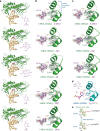
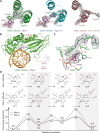
Fig. 1
Fig. 1. Tautomerization-dependent recognition of diverse substrates by hNEIL1.
a Overall views of hNEIL1(K242) bound to double-strand DNAs containing a 5-OHU, Sp1, (S)-Gh, DHT, or DHU. The chemical structures of individual damaged bases are shown in purple. b Zoom-in views of the interaction mode for hNEIL1(K242) in complex with various damaged bases. The lesion recognition loop located between αG and αH is indicated. The 2Fo−Fc simulated-annealing composite omit electron density map is shown in gray mesh and contoured at 1.0σ for both damaged bases and key loop residues. The gold dashed line represents hydrogen bond interaction. The dashed curve stands for an unbuildable short region of the lesion recognition loop due to ambiguous electron density. c The 242-in loop conformation was again observed in the crystal structure of a naturally existing variant hNEIL1(R242) in complex with 5-OHU, Sp1, and (S)-Gh, respectively. d The previously reported hNEIL1(R242)-Tg structure (PDB code: 5ITY) also shows a 242-in conformation, consistent with the all above described hNEIL1–DNA complexes. e The tautomerization-dependent hydrogen bonding between the flipped bases and residue 242 of hNEIL1. The base tautomers are denoted with the lactam (upper) and lactim (lower) forms.
Fig. 2
Fig. 2. Characterization of a newfound hNEIL1–DNA interaction mode.
a Crystal structure of hNEIL1(R242) bound to DHU (left) reveals a distinct conformation of lesion recognition loop, compared with the hNEIL1(R242)-Tg structure (middle) or the hNEIL1(R242)-APO structure (right). In the DHU-bound structure, Arg242 occupies the same position which is originally taken by Tyr244 of the Tg-containing structure, while Tyr244 stacks against the flipped DHU base. In addition, Arg242 of the APO structure is placed far away from the damaged bases and fully exposed to the solvent. The gray 2Fo−Fc simulated-annealing composite omit electron density map is contoured at 1.0σ for both damaged bases and lesion recognition loop. b Overall structure of hNEIL1(R242) bound to dsDNA-containing FDHU. The chemical structure of FDHU deoxynucleotide is shown in purple and residue 242 is colored in red. c Zoom-in view of the interactions between hNEIL1(R242) and FDHU. The brown dashed lines represent hydrogen bond interactions and red ball stands for water molecules. d QM/MM calculations reveal that the free-energy profile of base cleavage reaction pathway for 244-in conformation displays an extremely high energy barrier, comparing to a more reasonable 242-in conformation.
Fig. 3
Fig. 3. A proposed nucleobase-recognition model of hNEIL1 by computational and structural evidences.
a Adaptive MD simulations reveal the existence of three conformational states of hNEIL1 when interacting with the substrate DNA (DHU) with relatively enriched population. lmA local minimum A, lmB local minimum B, lmC local minimum C. The dots superimposed are randomly selected MD configurations from each metastable conformation and colored accordingly. b The loop conformations of snapshots from adaptive molecular dynamics simulations are in good coincidence with those observed in the crystal structures. c Substrate-recognition model of hNEIL1. Upon encountering the DNA substrates, the lesion recognition loop of hNEIL1 shifts towards the flipped bases from the originally preferred apo conformation (Encounter state). Once the tautomerization-dependent hydrogen bond interaction between damaged base and residue 242 is established, the substrate would be activated (Activated state) and permitted to enter the catalysis cycle. Alternatively, if the base does not pass the chemical check, the loop would transit to a 244-in conformation (Quarantine state), temporally restricting the nucleobase from catalysis via competing with the activated state. d The relative dominance of the quarantine state and activated state depends on the tug of war between their thermodynamic stability. ΔG denotes free energy difference quantifying the relative stability of the activated state over the quarantine state. ΔG can be decomposed into two terms, ΔGconf (Supplementary Fig. 9) and ΔGchem, respectively. The gold dashed line represents a process involving conformational transition and the blue solid line means a chemical transition.
Fig. 4
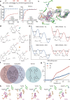
Fig. 4. Both key loop residues and base per se contribute to the state transition.
a The amino acid sequence of lesion recognition loop. Three key residues at position 242, 244 and 249 are indicated. b Biochemical assays show that hNEIL1(Y244R) is capable of rescuing the glycosylase activities of 242 mutants on DHU. Values represent mean ± s.e.m. (n = 3). See Supplementary Table 3 for calculated rate constants. c Zoom-in view of the interactions between FDHU and hNEIL1(Y244R). The simulated-annealing 2Fo−Fc composite omit electron density map is shown in gray mesh and contoured at 1.0σ. d An additional stable protonation state (“Tau-K2”) can be stabilized by Lys242, but bot by Arg242, to help retain the flipped base in the activated state (illustrated with DHU here). e The free energy profile of state transition for hNEIL1(K242) and hNEIL1(R242) bound to duplex DNA containing DHU or Tg. The calculated ΔGconf and ΔGchem values are determined in kcal/mol. Q state denotes quarantine state and A state stands for activated state. f The flipped nucleobases can be categorized by their chemical properties including non-planarity and hydrophilicity. Loss of aromaticity and increase of hydrophilicity of bases would facilitate the formation of a relatively stable activated state. g The number of desolvated water molecules (ΔNwater) when the loop transits from 242-in to 244-in conformation as a function of the distance from the base computed by MD simulations. h The observed solvent environments in the vicinity of the flipped bases within the crystal structures. Water molecules within 5 Å distance of the damaged base are shown here. The annealed difference Fo−Fc electron density map is shown in orange mesh and contoured at 2.5σ.
Fig. 5
Fig. 5. The interrogation of normal base by hNEIL1.
a The free energy profile of state transition for hNEIL1(R242) and hNEIL1(K242) bound to a mismatched dT. b Overall structure of hNEIL1(R242) bound to double-strand DNA containing a T:C mismatch. The flipped dT is colored with purple and Arg242 is shown in red. c Zoom-in view of the interactions between dT and hNEIL1(R242). A quarantine state similar to that identified in the crystal structures of hNEIL1(R242) in complex with (F)DHU or DHT is also observed here. The 2Fo−Fc simulated-annealing composite omit electron density map is shown in gray mesh and contoured at 1.0σ. d An electrophoretic assay shows the glycosylase activities of unedited hNEIL1(K242) and edited hNEIL1(R242) on a T:C mismatch. The experiment was repeated three times independently with similar results. Source data are provided as a Source Data file. Detailed quantitative results can be found in Supplementary Fig. 13.

References

    1. Lindahl T. Instability and decay of the primary structure of DNA. Nature. 1993;362:709–715. doi: 10.1038/362709a0. - DOI - PubMed
    1. Hoeijmakers JH. DNA damage, aging, and cancer. N. Engl. J. Med. 2009;361:1475–1485. doi: 10.1056/NEJMra0804615. - DOI - PubMed
    1. Roos WP, Thomas AD, Kaina B. DNA damage and the balance between survival and death in cancer biology. Nat. Rev. Cancer. 2016;16:20–33. doi: 10.1038/nrc.2015.2. - DOI - PubMed
    1. Krokan, H. E. & Bjoras, M. Base excision repair. CSH Perspect. Biol.5, a012583 (2013). - PMC - PubMed
    1. David SS, O’Shea VL, Kundu S. Base-excision repair of oxidative DNA damage. Nature. 2007;447:941–950. doi: 10.1038/nature05978. - DOI - PMC - PubMed

Publication types

LinkOut - more resources